
Dr. Prerna Sahrawat
Contributing Dental Clinician
Intraoral Scanner Technology Explained
Digital Impression Core Concepts
The shift to digital workflows is accelerating in modern dental practices. These advanced devices are at the heart of this transformation, replacing traditional methods with precise, efficient digital data collection.
Table of Contents
Core Scanning Technologies Explained
- Confocal microscopy captures sharply focused points for high-detail images.
- Triangulation projects patterns and calculates depth from their deformation.
- Active wavefront sampling analyzes how light reflects off tooth surfaces.
- Structured light projects grids or lines to precisely map surface topography.
- These methods progressively build a 3D model point by point.
- Each technology has unique benefits for scanning speed and surface detail.
HOW DIGITAL SCANNING WORKS
The wand projects a safe, patterned light source onto the teeth and gingiva.
A high-speed sensor captures thousands of images per second from multiple angles.
Software algorithms stitch the images together into a precise and final 3D model.
The completed model is exported as an STL or PLY file for lab use.
Speed, Accuracy, and Colour
- Scan speed is measured in frames per second (FPS) or by arch capture time.
- Higher speeds allow for faster and more comfortable data acquisition for patients.
- Accuracy is how true the digital model is to the actual patient anatomy.
- Trueness measures closeness to the object's real, physical dimensions.
- Precision measures the repeatability of multiple scans on the same object.
- Colour rendering helps differentiate between tissue types and restoration margins.
Open vs. Closed Systems
- Open systems export universal file formats like STL, PLY, and OBJ.
- This provides maximum flexibility to work with any lab or design software.
- Closed systems use proprietary formats, restricting you to one ecosystem.
- These often provide a more seamless and integrated chairside workflow.
- Consider your long-term goals for lab partnerships and in-house milling.
- Analyze subscription fees and software update costs for each model.
Open Systems
- ✅ Maximum lab and equipment flexibility
- ✅ No proprietary file restrictions
- ✅ Often lower long-term costs
- ❌ May require more initial setup
Closed Systems
- ✅ Seamless hardware and software integration
- ✅ Simplified and guided clinical workflows
- ✅ Dedicated, single-source customer support
- ❌ Limits future lab and equipment choices
Beyond Digital Impressions
- Create digital patient records to monitor wear, erosion, and tooth movement.
- Plan implant placements with surgical guides for enhanced precision and safety.
- Fabricate clear aligners and orthodontic appliances from accurate models.
- Design and mill same-day crowns, inlays, and onlays with chairside systems.
- Improve patient communication with realistic 3D visualizations of their mouth.
- Collaborate seamlessly with dental labs by sending digital files instantly.
EXPANDED CLINICAL WORKFLOWS
Design crowns, bridges, inlays, onlays, and veneers with high accuracy.
Plan implant positions and fabricate custom surgical guides for placement.
Create models for clear aligners and for tracking treatment progress.
Track tooth wear and gingival changes over time with scan overlays.
Maximize Your Scan Data
For wear analysis, take a baseline full-arch scan for every new patient. This digital record, stored in under 5 minutes, becomes an invaluable tool for monitoring changes over many years.
Frequently Asked Questions
Most clinicians become proficient with basic scanning in just a few sessions. Achieving expert speed and efficiency for a full arch scan, typically under 60 seconds, can take about 20-30 practice scans. Most manufacturers provide comprehensive training and support to shorten this learning period significantly.
Yes, with proper technique. Achieving accurate subgingival margin capture requires excellent tissue retraction and moisture control, similar to traditional methods. Some devices feature advanced software algorithms that help highlight the margin line, but clinical skill remains paramount, often requiring a dual-cord technique for best results.
Scan powder was used on older systems to create a non-reflective, uniform surface for easier data capture. However, over 95% of modern devices are 'powder-free,' using advanced optics and software that can accurately read shiny or translucent enamel without any surface coating, which greatly simplifies the workflow.
An STL (Standard Tessellation Language) file is the most common format, containing only the geometric surface data of the 3D model. A PLY (Polygon File Format) file is a more advanced format that includes both the geometry and the full-colour texture data, providing a more realistic and diagnostically useful model.
The physical footprint is minimal. Most systems consist of a wand, which is about the size of a large electric toothbrush, and a laptop or a dedicated cart with a monitor. The cart-based systems typically require about 4 square feet of floor space, while laptop versions are completely portable between operatories.
Written by
Dr. Prerna Sahrawat
Contributing Dental Clinician
Dr. Prerna Sahrawat is a dentist with over 4 years of experience in clinical dentistry and dental content writing. She is passionate about simplifying complex dental concepts into practical, evidence-based content that helps dentists make informed clinical and purchasing decisions while staying updated with the latest advancements in dentistry.
Keep reading
Browse all →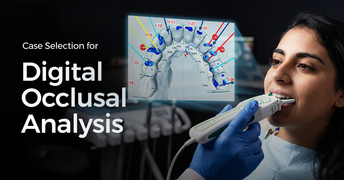
Case Selection for Digital Occlusal Analysis
Case Selection for Digital Occlusal AnalysisA Clinical Decision Guide A digital occlusal analysis system provides objective, dynamic data on bite timing and for

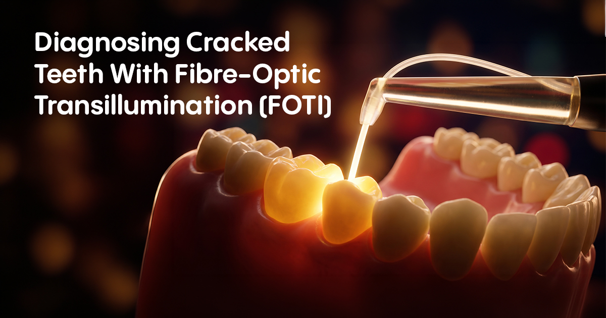
Diagnosing Cracked Teeth With Fibre-Optic Transillumination
Diagnosing Cracked Teeth With Fibre-Optic Transillumination (FOTI)A Clinical Selection Guide Fibre-optic transillumination (FOTI) is a diagnostic method using a
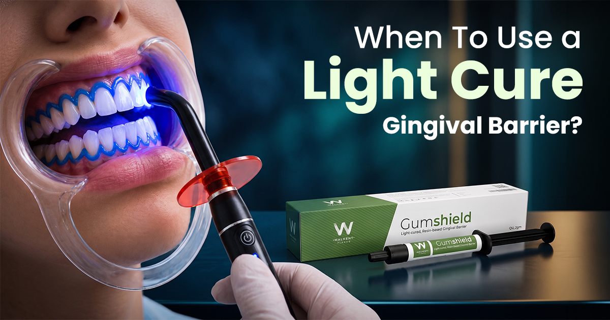
When To Use a Light Cure Gingival Barrier?
When To Use a Light Cure Gingival Barrier?A Clinical Selection Guide. A liquid gingival barrier is a light-curable resin used to isolate and protect soft tissue