
Dr. Bhavishya Arora
Chief Dental Editor
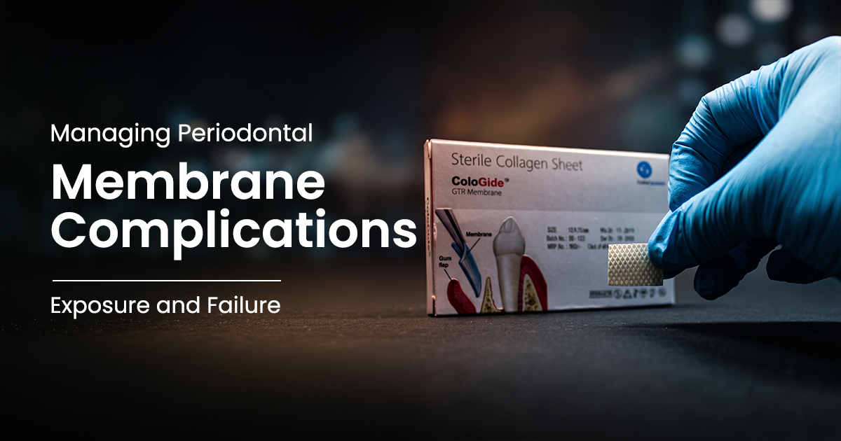
Managing Exposed GTR Membranes: Clinical Strategies
Optimizing Regenerative Outcomes
Effective Guided Tissue Regeneration (GTR) membrane management requires primary soft tissue closure over the barrier to prevent bacterial contamination and ensure predictable bone regeneration. Premature exposure, the most common post-surgical complication, directly compromises regenerative potential.
Table of Contents
Causes of GTR Membrane Exposure
GTR membrane exposure in periodontics primarily results from inadequate soft tissue coverage and excessive flap tension during suturing, leading to wound dehiscence.
Tension-free, passive primary closure is crucial for guided tissue regeneration success. Overly stretched mucoperiosteal flaps compromise blood supply, risking necrosis and dehiscence within 7-14 days. Improper flap design, such as inadequate releasing incisions or periosteal release failure, creates tension, pulling flap margins apart. Preventing this requires appropriate periodontic products, suture materials, and instruments.
- Flap Tension: Insufficient periosteal releasing incisions cause flap retraction and marginal ischemia.
- Improper Suturing: Sutures too close to the margin or tied excessively can cause tissue necrosis.
- Membrane Instability: Poor adaptation or stabilization with tacks creates pressure points under the flap.
- Patient Factors: Poor oral hygiene, smoking, or trauma disrupts healing, leading to dehiscence.
KEY FACTORS LEADING TO MEMBRANE EXPOSURE
Inadequate flap release compromises blood supply, causing marginal necrosis and dehiscence.
Improper suture placement or excessive tightness can strangulate tissue, causing wound breakdown.
Smoking and poor oral hygiene introduce contaminants, impairing initial healing.
Impact of Early Exposure on Guided Tissue Regeneration
Early guided tissue regeneration membrane exposure severely compromises regenerative outcomes by introducing bacterial contamination, causing inflammation and incomplete bone fill. This converts a protected regenerative environment into an infection-susceptible site.
Exposed membrane surfaces rapidly colonize with oral bacteria, forming a biofilm within 24-48 hours. This triggers a persistent inflammatory response, down-regulating osteoblastic activity and promoting fibrous tissue instead of new bone. Compromise extent relates directly to exposure duration and size; exposures >3 mm often yield minimal to no bone gain.
- Bacterial Contamination: Oral pathogens colonize the membrane, leading to local infection and hindering cell proliferation.
- Inflammatory Response: Chronic inflammation prevents osteoprogenitor cell migration and differentiation into the graft.
- Soft Tissue Invagination: Gingival epithelium can migrate along the exposed membrane, preventing clot stabilization and space maintenance.
- Reduced Bone Fill: Clinical outcomes show a direct negative correlation between exposure area and defect fill percentage.
Critical Exposure Window
Exposure within the first 2-4 weeks post-surgery is critical. The organizing blood clot is highly vulnerable to bacterial contamination, potentially leading to complete regenerative procedure failure.
Strategies to Mitigate GTR Complications
Mitigating GTR membrane complications involves meticulous surgical technique for passive primary closure and diligent post-operative care. Proactive management of minor exposures often salvages regenerative outcomes.
Surgical precision is the first defense: design flaps for tension-free closure, perform adequate periosteal releasing incisions, and use appropriate suturing (e.g., vertical/horizontal mattress). For minor exposures (<3 mm), a conservative approach is best. Instruct patients on gentle site care and apply 0.12% chlorhexidine gluconate gel twice daily to control bacterial load, allowing secondary intention healing.
- Achieve Passive Closure: Ensure the flap passively covers the membrane completely before suturing.
- Use Cross-Linked Membranes: Consider membranes with longer resorption profiles in high-risk areas for better bacterial resistance.
- Implement a CHX Regimen: For small exposures, prescribe 0.12% chlorhexidine rinse for 2-4 weeks to control microbial plaque.
- Avoid Probing: Instruct patients and staff to avoid mechanical probing or disrupting the exposed site.
Post-Exposure Clinical Checklist
Measure dimensions. Exposures <3 mm are often conservatively manageable.
Evaluate for purulent exudate, swelling, or erythema, which may necessitate membrane removal.
Prescribe 0.12% CHX rinse twice daily to reduce local bacterial load.
Educate patient on gentle brushing of adjacent teeth, avoiding direct surgical site contact.
Monitor weekly for the first month to track granulation and ensure exposure isn't enlarging.
Indications for GTR Membrane Removal
GTR membrane removal is indicated for significant exposure with clinical infection signs, such as purulent exudate, persistent swelling, or pain. This resolves infection and prevents further graft and bone destruction.
While small, asymptomatic exposures are conservatively manageable, large exposures (>3-4 mm) or any with frank suppuration create an unresolvable septic condition. The membrane acts as a foreign body, perpetuating infection and preventing healing. Here, the priority shifts from regeneration to infection control. Prompt removal, using high-quality periodontal instruments, salvages underlying augmentation.
- Persistent Infection: Purulent discharge, fistula, or abscess unresolved by antimicrobial rinses.
- Extensive Exposure: Dehiscence exceeding 30-50% of the membrane, making conservative management futile.
- Patient Discomfort: Significant pain, discomfort, or foul taste attributable to the surgical site.
- Soft Tissue Proliferation: Extensive soft tissue invagination under the membrane, indicating complete loss of regenerative space.
GTR Membrane: Retain or Remove?
Long-Term Prognosis After Membrane Exposure
Long-term prognosis after GTR membrane exposure varies, depending on exposure size, duration, and intervention timing. Small, well-managed exposures may yield acceptable results; large, infected ones often lead to compromised or failed regeneration.
Effectively managed small exposures with antimicrobial rinses, healing by secondary intention, may result in slightly less than planned bone gain. However, premature membrane removal due to infection typically yields poor regenerative outcomes, with healing by dense connective tissue instead of bone. Such cases may require secondary augmentation after at least 6 months. Careful GTR membrane management is thus key to prognosis.
- Minor Exposure (<3 mm): Often yields 50-70% of anticipated bone gain if infection-free.
- Major Exposure (>3 mm): Typically yields less than 25% of expected bone gain, often with soft tissue invagination.
- Premature Removal (Infection): Poor prognosis; regeneration often fails completely. Resolving infection becomes the primary goal.
- Secondary Procedures: Failed GTR sites may be candidates for re-treatment after a 6-9 month healing interval for soft tissue maturation.
Conservative Management
- Minimally invasive, preserves graft
- Can salvage partial regeneration
- Risk of infection remains
- Requires high patient compliance
Surgical Removal
- Definitive infection control
- Allows for soft tissue healing
- Sacrifices the regenerative potential
- May require a second surgery later
Frequently Asked Questions
Ideally, a GTR membrane should remain submerged for 4-6 weeks to allow initial angiogenesis and bone graft consolidation. Exposure before this critical window severely disrupts healing by introducing bacteria and preventing stable blood clot formation, foundational to successful GTR membrane management.
Resorbable membranes (e.g., collagen) may heal by secondary intention with small exposures but degrade quickly when contaminated. Non-resorbable membranes (e.g., d-PTFE) don't degrade but are highly prone to bacterial wicking if exposed, almost always requiring removal to resolve infection.
For minor GTR membrane exposure, weekly follow-ups are recommended for the first 3-4 weeks. This monitors infection signs, assesses exposure stability, and reinforces oral hygiene, ensuring prompt addressing of negative changes.
A periosteal releasing incision combined with a double-layer suturing technique is highly effective. An internal horizontal mattress suture approximates flap margins without tension, then simple interrupted sutures achieve precise wound edge eversion and a primary seal.
Yes, trimming the exposed, contaminated membrane portion is a viable strategy for exposures >3-4 mm with no frank infection. After local anesthesia, the supracrestal membrane part is carefully excised, allowing soft tissue granulation over the remaining submerged portion.
Written by
Dr. Bhavishya Arora
BDS, MDSChief Dental Editor
A contributing clinician at Dentalkart Blogs, Dr. Bhavishya is a compassionate, detail-driven dentist known for thoughtful patient care, clear communication, and a calm chair-side manner that puts patients at ease. Beyond the clinic, she leads product content and category initiatives at Dentalkart India's largest dental marketplace where she bridges clinical understanding with commercial insight to help dentists make better-informed product choices.
Keep reading
Browse all →
Indications for Periodontal Dressing Placement
Indications for Periodontal Dressing PlacementA Clinical Case Selection Guide A periodontal dressing material is a protective pack placed over a surgical site t

Indications for Periodontal Fiber Splinting
Indications for Periodontal Fiber SplintingA Clinical Case Selection Guide Periodontal splinting is indicated for stabilizing teeth with increasing mobility (Mi
Preventing Burnished Calculus with Gracey Curettes
Preventing Burnished Calculus with Gracey CurettesA Subgingival Scaling Protocol Burnished calculus is a residual deposit smoothed by an incorrectly used curett