
Dr.Yukti
Dental Content Contributor
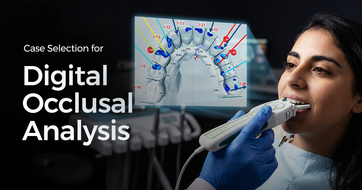
Case Selection for Digital Occlusal Analysis
A Clinical Decision Guide
A digital occlusal analysis system provides objective, dynamic data on bite timing and force, essential for complex restorative, implant, and TMD cases where articulating paper's qualitative marks are insufficient. Precision diagnoses hidden interferences, ensuring long-term treatment success.
Table of Contents
Clinical Signs Demanding Digital Analysis
Clinical signs indicating a need for digital analysis include unexplained ceramic fractures, persistent patient complaints of a 'high bite' despite adjustments, and fremitus on specific teeth. These symptoms suggest underlying occlusal discrepancies related to force and timing that static marks from articulating paper cannot reveal.
Digital occlusal analysis quantifies contact sequence and force percentage, pinpointing premature contacts (e.g., one receiving 40% of total bite force milliseconds before others). This objective data diagnoses issues beyond clinical guesswork, leading to targeted, effective adjustments.
- Unexplained Prosthesis Failure: Recurrent chipping or fracture of ceramic restorations (e.g., veneers, all-ceramic crowns) with no other apparent etiology.
- Persistent Hyperocclusion: Patients reporting persistent discomfort or 'the bite is off' after conventional adjustments.
- Wear Facets & Abfractions: Atypical wear patterns or non-carious cervical lesions correlating with suspected high occlusal loads.
- Fremitus: Palpable or visible tooth vibration (fremitus) upon clenching, indicating excessive, focused occlusal force requiring quantification.
- Post-Orthodontic Instability: Unexplained minor tooth movement or relapse post-orthodontic treatment, suggesting unresolved occlusal interferences.
Digital Analysis Indication Checklist
Repeated crown/veneer chipping despite seemingly correct occlusion.
Consistent 'high bite' complaint not located by articulating paper.
Specific tooth (often anterior) vibrates upon clenching.
Full-mouth rehabilitation or multiple implant-supported units required.
Prioritizing Prosthodontic Cases for Digital Occlusion
Full-mouth rehabilitations, multiple implant prostheses, and anterior aesthetic cases benefit most from digital occlusal analysis to manage complex force distribution and ensure predictable outcomes. Balanced, simultaneous, harmonious occlusion is paramount for restoration longevity and stomatognathic system health.
Implants lack periodontal ligament, eliminating natural proprioceptive feedback against overloads. A digital system is critical to ensure no single implant bears disproportionate load, targeting an initial force distribution under 15-20% per implant site in full-arch cases. For anterior veneers, it allows precise calibration of canine guidance for immediate posterior disclusion (<0.5 mm lateral movement), protecting fragile porcelain.
- Full-Arch Implant Prostheses: Verifies passive, stable occlusal schemes and distributes load evenly across implants to prevent screw loosening or component fracture.
- Complex Crown & Bridge: For cases with >4 units, especially midline-crossing, ensuring occlusal design does not introduce iatrogenic interferences.
- Anterior Aesthetic Restorations: Precisely engineers canine guidance and anterior disclusion, protecting porcelain veneers from chipping during excursive/protrusive movements.
- Digital Smile Design (DSD) Cases: Functionally validates the new occlusal vertical dimension (OVD) and envelope of function proposed in aesthetic/functional wax-ups.
Prosthodontic Case Selection Matrix
TMD Patients: When is Digital Analysis Indicated?
No, digital analysis isn't routine for all TMD patients; it's most valuable for those with a clear occlusal component, such as myalgia linked to specific prematurities. It aids myogenous TMD diagnosis but doesn't diagnose intra-articular disorders (e.g., disc displacement), which require imaging and clinical evaluation.
For patients with myofascial pain, digital occlusal analysis identifies premature contacts triggering muscle splinting and hyperactivity. Pinpointing/eliminating this occlusal trigger often achieves significant symptom relief. The data aims to create an occlusion minimizing muscular effort, often via Disclusion Time Reduction (DTR) therapy.
- Diagnostic Splint Therapy: Use before/after splint delivery to objectively measure occlusal contact changes and guide adjustments for a therapeutic position.
- Suspected Occlusal Triggers: Indicated when TMD symptoms begin after a new restoration, suggesting iatrogenic interference.
- Pre-Prosthetic Assessment: Crucial for TMD patients needing extensive restorative work, to establish a stable, comfortable occlusal foundation.
- Documenting Masticatory Dynamics: Records and analyzes occlusal patterns in patients with chronic muscle soreness, bruxism, or clenching habits.
Clinical Exam Only
- Accessible and requires no special equipment.
- Effective for diagnosing most intra-articular issues (e.g., clicking, crepitus).
- Relies on subjective patient feedback for bite assessment.
- Cannot quantify occlusal force or timing of contacts.
With Digital Analysis
- Provides objective, repeatable data on force and time.
- Identifies specific premature contacts that can trigger myalgia.
- Higher initial investment in equipment and training.
- Data interpretation requires specific expertise to be clinically useful.
Defining the Role of Traditional Articulating Paper
Articulating paper remains sufficient for routine, single-unit restorations, interproximal contact checks, and initial gross occlusal adjustments where qualitative contact location is the primary goal. It's a fast, inexpensive, indispensable tool for most daily dental procedures in a high-volume Indian practice.
Articulating paper's fundamental limitation is that ink mark size/intensity doesn't reliably correlate with occlusal force. A broad, dark mark can result from a low-force, flat cusp fossa contact, while a tiny, faint mark could be a high-force premature contact on a cusp tip. It shows where teeth touch, not how or when with specific force levels.
- Single-Unit Restorations: Perfect for adjusting single Class I composites or PFM crowns in an existing, stable occlusal scheme.
- Initial Gross Adjustments: Quickly identifies highest spots on a new prosthesis before finer, more detailed adjustments.
- Denture Adjustments: Checks contacts on acrylic denture teeth, where achieving bilateral balance is more critical than precise force measurement.
- Provisional Restorations: Ensures temporary crowns aren't in hyperocclusion and are comfortable pending final restoration delivery.
- Post-Debonding Check: Quick verification after orthodontic bracket removal to ensure no residual bonding agent creates interference.
Maximizing Articulating Paper Accuracy
Always ensure the teeth are completely dry before marking to prevent smudging. Use a dual-color, thin articulating paper (e.g., 40-micron thickness) to mark centric contacts in one color and excursive interferences in another for a more effective, albeit qualitative, diagnosis.
Conclusion: A Precision-Driven Approach
Digital occlusal analysis transitions from qualitative assumptions to quantitative data for complex cases. Indian clinicians must decide when to augment articulating paper with dynamic force/timing information to prevent failures in high-stakes restorative and TMD treatments. This elevates clinical predictability, enhancing long-term patient satisfaction by addressing root occlusal problems.
- Full-Arch Cases: Mandate digital analysis for multi-unit implant prostheses to balance forces (<20% per site), preventing component failure.
- Diagnostic Clarity: Use digital analysis to investigate repeated ceramic fractures or phantom high spots when articulating paper shows no obvious errors.
- Myogenous TMD: Reserve for TMD cases with a suspected occlusal trigger, identifying/eliminating specific premature contacts causing muscle hyperactivity.
- Routine Work: Rely on high-quality, dual-color articulating paper (around 40 microns) for routine single-unit adjustments in stable occlusions.
Frequently Asked Questions
A digital occlusal analysis system uses an intraoral sensor to measure and record relative force and timing of tooth contacts during biting. Unlike articulating paper (location only), this technology provides objective, dynamic 2D/3D data, revealing otherwise invisible premature contacts and force imbalances.
The main difference is dynamic versus static information. A traditional articulator simulates jaw movements based on static records (e.g., bite registrations), showing potential contact locations mechanically. Digital occlusal analysis records the patient's actual, real-time, muscle-driven occlusal function, measuring precise timing and force of every contact, which an articulator cannot.
For full-mouth rehabilitation, digital analysis should be used at key stages: initially for diagnosis, after provisionals to refine the occlusal scheme, and at final insertion to verify definitive prostheses. It is also valuable during follow-up (1 and 6 months post-treatment) to check occlusal settling and ensure long-term stability.
While any dentist can operate the hardware, interpreting data effectively requires specific training. Manufacturers often offer certification for concepts like Disclusion Time Reduction (DTR) and force-vs-time graphs. This knowledge is crucial to translate raw data into clinically relevant, precise adjustments, moving beyond identifying high spots to understanding the entire occlusal cycle.
This can arise from several factors. First, ensure correct sensor positioning and that the patient bit in their habitual centric occlusion, not a convenience bite. Second, verify precise, non-aggressive adjustments, as this can shift forces elsewhere. Finally, consider non-occlusal causes like pulpal inflammation or referred muscle pain, as the digital occlusal analysis system only assesses the bite.
Written by
Dr.Yukti
Dental Content Contributor
Dr. Yukti Jain is a BDS-qualified dental professional and Product Specialist at Dentalkart with expertise in dental materials, equipment, and clinical innovations. Passionate about evidence-based dentistry, she creates insightful, research-driven content that simplifies complex topics and empowers dental professionals to make informed clinical and purchasing decisions.
Keep reading
Browse all →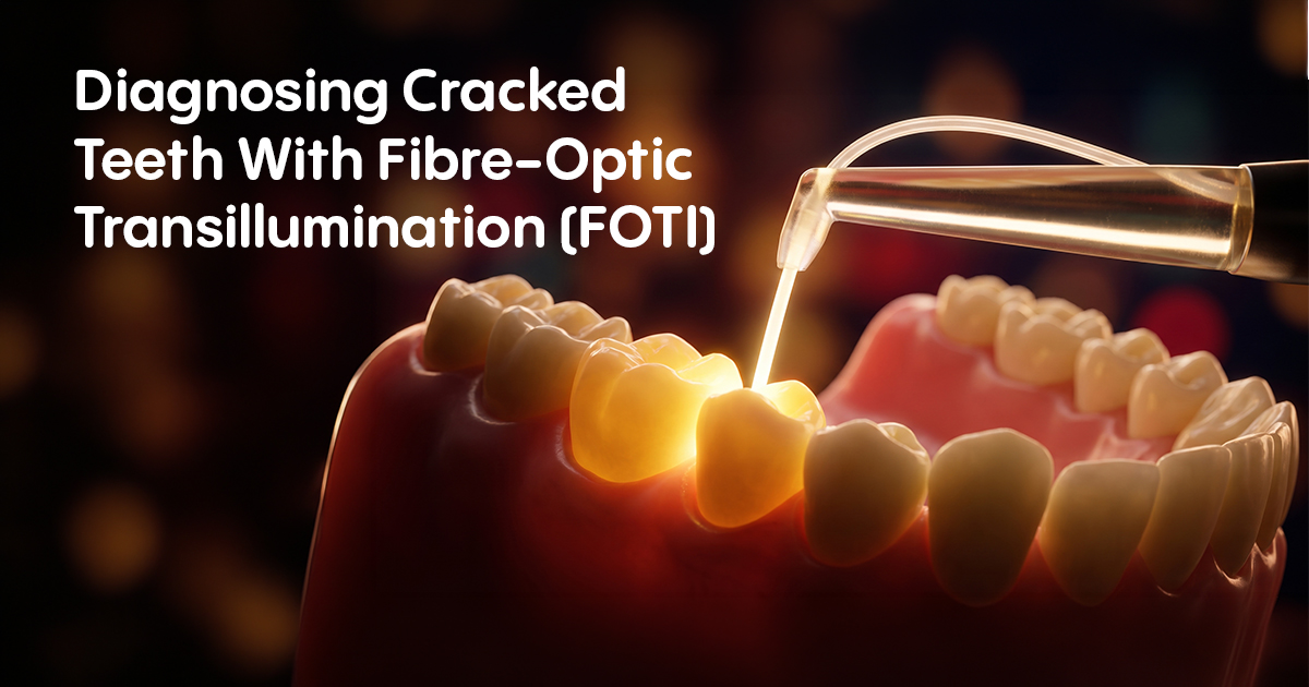
Diagnosing Cracked Teeth With Fibre-Optic Transillumination
Diagnosing Cracked Teeth With Fibre-Optic Transillumination (FOTI)A Clinical Selection Guide Fibre-optic transillumination (FOTI) is a diagnostic method using a
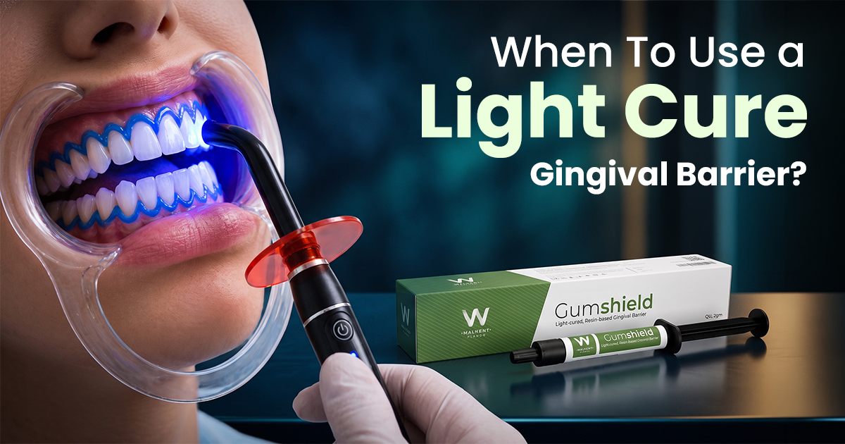
When To Use a Light Cure Gingival Barrier?
When To Use a Light Cure Gingival Barrier?A Clinical Selection Guide. A liquid gingival barrier is a light-curable resin used to isolate and protect soft tissue
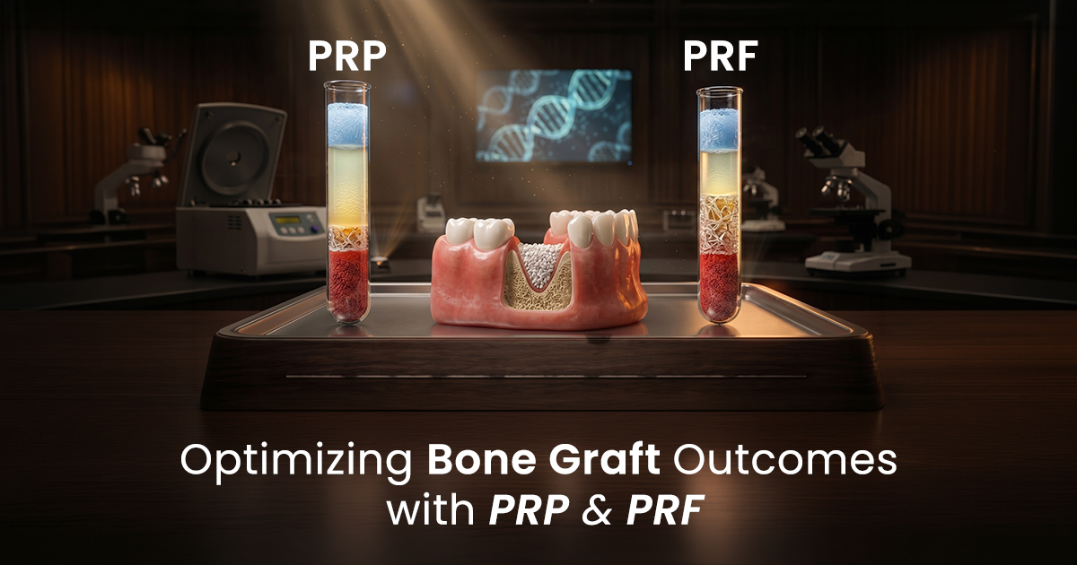
Optimizing Bone Graft Outcomes with PRF and PRP — Enhancing Healing in Oral Surgery
Optimizing Bone Graft Outcomes with PRF and PRPEnhancing Healing in Oral Surgery PRF and PRP are autologous blood concentrates that deliver high concentrations
