
Dr.Yukti
Dental Content Contributor
Diagnosing Cracked Teeth With Fibre-Optic Transillumination (FOTI)
A Clinical Selection Guide
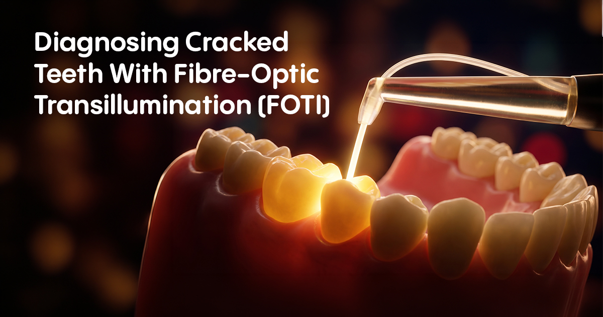
Fibre-optic transillumination (FOTI) is a diagnostic method using a high-intensity light to visualize fracture lines within a tooth structure. Its correct application is crucial for differentiating symptomatic cracks from benign craze lines, guiding treatment and avoiding unnecessary endodontic procedures.
Table of Contents
Which Symptoms Indicate a Need for FOTI?
FOTI is indicated when a patient presents with sharp, fleeting pain on biting or release, which cannot be attributed to caries, defective restorations, or periodontal issues. This classic cracked tooth symptom, especially when localized to a specific cusp but difficult to pinpoint visually, is a primary trigger for using a dental transilluminator.
The underlying mechanism is cuspal flexure during mastication, which irritates the pulp. The pain upon release of biting pressure is pathognomonic, as the crack segments spring back into position. A dental transilluminator works by passing a focused beam of light through the tooth; a fracture line, having a different refractive index, disrupts this light path and appears as a distinct dark line, confirming the source of the elusive pain.
- Sharp pain on biting: Especially when chewing granular foods like grains or nuts.
- Pain on release of pressure: A key indicator, as the fractured segments move.
- Localized thermal sensitivity: Particularly to cold, with no other identifiable cause.
- Inconclusive radiographs: Fractures, especially in the mesiodistal plane, are often invisible on 2D X-rays.
- No visible evidence: The tooth may appear clinically sound with no caries or restoration failure.
Clinical Checklist: When to Use FOTI
This is the most common and reliable symptom of cracked tooth syndrome.
FOTI helps isolate the specific tooth and cusp involved.
Transillumination can reveal fractures that are otherwise undetectable.
Use FOTI to confirm the presence and extent of the fracture line on the suspected cusp.
FOTI vs. Methylene Blue Dye for Crack Detection
The primary difference between FOTI and methylene blue dye is that FOTI is a non-invasive, real-time diagnostic aid, while dye staining is an invasive technique that requires tooth preparation and can cause permanent discoloration. FOTI is the preferred initial method for vital teeth as it provides immediate chairside information without altering the tooth structure.
Methylene blue dye works via capillary action, seeping into the fracture line and making it visible upon removal of the excess. While effective, it can permanently stain dentin, complicating aesthetic outcomes. A high-intensity LED dental transilluminator, by contrast, relies purely on light physics and is completely reversible, making it a superior choice for initial assessment and patient communication.
- Reversibility: FOTI is completely non-invasive and reversible; dye staining is not.
- Speed: Transillumination provides an instant result, whereas dye requires application, waiting, and cleanup.
- Scope: FOTI can help visualize cracks through intact marginal ridges, while dye is most effective on exposed surfaces after restoration removal.
- Patient Acceptance: Patients readily accept a light-based examination over the application of a strong, potentially staining chemical.
| Attribute | FOTI (Transillumination) | Methylene Blue Dye |
|---|---|---|
| Invasiveness | Non-invasive best | Invasive (can stain) |
| Diagnostic Speed | Immediate | Delayed (requires application time) |
| Best Use Case | Initial diagnosis on vital teeth | Confirming cracks on prepared surfaces |
| Subgingival Detection | Poor | Can be effective value |
| Equipment Needed | Fibre-optic light source | Dye, micro-applicators, cleaning pellets |
What Are the Limitations of FOTI Diagnosis?
FOTI diagnosis is most limited by the presence of certain dental materials and the location of the crack itself. Full coverage crowns, particularly PFM or full metal, completely block light transmission, rendering FOTI useless for detecting cracks underneath them. Similarly, large, multi-surface composite restorations can scatter the light, obscuring underlying fracture lines.
The technique's efficacy diminishes significantly for cracks that extend deep subgingivally or are located entirely within the root structure. The light from a dental transilluminator cannot adequately penetrate bone and soft tissue to illuminate a vertical root fracture. Therefore, while excellent for coronal cracks in minimally restored teeth, FOTI is not a definitive tool for assessing restoratively complex teeth or suspected root fractures.
- Restorative Materials: Opaque materials like metal, PFM, and some ceramics will block light.
- Crack Location: Does not work for subgingival or radicular fractures.
- Tooth Discoloration: Intrinsic staining or pulpal necrosis can create dark areas that mimic cracks.
- Operator Skill: Correct angulation of the light source is critical for accurate interpretation.
Diagnostic Blind Spots: When Not to Rely on FOTI
Never use FOTI as the sole diagnostic tool for teeth with full coverage PFM or metal crowns. A negative FOTI result in such cases is meaningless. Suspected cracks under these restorations require removal of the crown and direct visual inspection, often supplemented with dye.
Differentiate Cracks from Craze Lines Using FOTI
Using a dental transilluminator, a crack is identified as a distinct dark line that stops or significantly impedes the transmission of light across the tooth, whereas a craze line typically does not. This is the fundamental difference in their FOTI presentation and is key to avoiding overtreatment.
Craze lines are superficial, incomplete fractures confined to the enamel and are almost always asymptomatic. Because they are shallow and often filled with organic material, they do not significantly alter the path of light. A true fracture, however, propagates into dentin, creating a physical separation that acts as a barrier to light. When transilluminating, sweep the light from buccal to lingual; a crack will appear as a sharp, dark border, while a craze line may be faintly visible or not at all.
- Light Transmission: A crack blocks light; a craze line usually transmits light.
- Termination Point: A fracture line will often terminate at a marginal ridge or extend onto the occlusal surface.
- Symptomatology: Correlate FOTI findings with clinical symptoms. Craze lines are asymptomatic.
- Location: Craze lines are common on anterior teeth and buccal surfaces of posterior teeth. Symptomatic cracks are most frequent in posterior teeth.
FOTI Interpretation: Crack vs. Craze Line
Conclusion: FOTI for Targeted Diagnosis
Fibre-optic transillumination is an indispensable, non-invasive chairside tool for the selective diagnosis of suspected cracked teeth. Its greatest value lies in confirming coronal fractures in vital, minimally restored posterior teeth where other signs are ambiguous. Integrating FOTI as a primary step after a positive bite test—but before resorting to invasive exploratory procedures—improves diagnostic accuracy, saves clinical time, and enhances patient trust.
- Ideal Indication: Use FOTI for sharp, fleeting pain on biting/release in minimally restored posterior teeth.
- Interpretation Rule: A true crack blocks light transmission, appearing as a dark line; a craze line does not.
- Key Limitation: Do not rely on FOTI for teeth with full-coverage metal or PFM crowns, or for suspected root fractures.
- Technique Tip: Always compare the suspected cusp with an adjacent healthy cusp to establish a baseline for normal light transmission.
Frequently Asked Questions
A dental transilluminator works by shining a high-intensity, focused beam of light through the coronal portion of a tooth. Healthy tooth structure transmits this light uniformly. However, structural defects like cracks, fractures, or caries have a different refractive index, causing them to scatter or block the light, making them visible as dark areas or lines.
Yes, FOTI is a useful adjunct for detecting interproximal caries, especially in posterior teeth. Carious dentin scatters light more than healthy dentin, causing the lesion to appear as a dark, shadowy area when illuminated from the buccal or lingual aspect. It is a valuable radiation-free alternative to bitewing radiographs for initial screening.
FOTI and CBCT serve different purposes. FOTI is an excellent, low-cost chairside tool for detecting coronal cracks but is ineffective for root fractures. A CBCT, while exposing the patient to radiation, is the gold standard for visualizing vertical root fractures and assessing the extent of fractures in relation to the alveolar bone. FOTI is for initial coronal assessment; CBCT is for complex radicular diagnosis.
While traditional FOTI uses a white light source, some modern devices offer different colours. An orange or red light (longer wavelength, around 625-740 nm) can sometimes enhance the visibility of cracks by reducing scattering from healthy tooth structure. However, a bright, focused white light remains the standard for most diagnostic applications.
A dedicated dental transilluminator is strongly recommended. While a curing light can be used in a pinch, its beam is typically wider, less collimated, and in the blue spectrum (around 450-495 nm), which is not ideal for diagnostic transillumination. A proper FOTI device provides a focused, high-intensity white light beam for superior clarity and diagnostic accuracy.
Written by
Dr.Yukti
Dental Content Contributor
Dr. Yukti Jain is a BDS-qualified dental professional and Product Specialist at Dentalkart with expertise in dental materials, equipment, and clinical innovations. Passionate about evidence-based dentistry, she creates insightful, research-driven content that simplifies complex topics and empowers dental professionals to make informed clinical and purchasing decisions.
Keep reading
Browse all →
Case Selection for Digital Occlusal Analysis
Case Selection for Digital Occlusal AnalysisA Clinical Decision Guide A digital occlusal analysis system provides objective, dynamic data on bite timing and for
When To Use a Light Cure Gingival Barrier?
When To Use a Light Cure Gingival Barrier?A Clinical Selection Guide. A liquid gingival barrier is a light-curable resin used to isolate and protect soft tissue
Optimizing Bone Graft Outcomes with PRF and PRP — Enhancing Healing in Oral Surgery
Optimizing Bone Graft Outcomes with PRF and PRPEnhancing Healing in Oral Surgery PRF and PRP are autologous blood concentrates that deliver high concentrations
