Dr.Bhavishya
Dental Content Contributor
Optimizing Bone Graft Outcomes with PRF and PRP
Enhancing Healing in Oral Surgery
PRF and PRP are autologous blood concentrates that deliver high concentrations of growth factors, accelerating healing in bone grafting. Leveraging these biologics improves surgical outcomes and patient recovery.
Table of Contents
PRF and PRP in Dental Bone Grafting
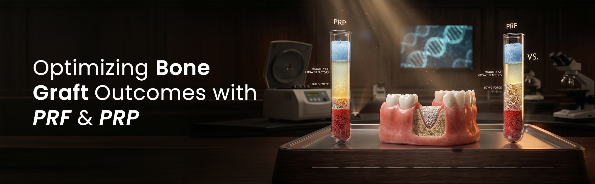
PRF and PRP are autologous biologics derived from a patient's blood, enhancing bone graft healing. They concentrate platelets and growth factors to stimulate tissue regeneration.
While both serve a similar purpose, their preparation and properties differ. PRP, a first-generation concentrate, requires anticoagulants and a two-step centrifugation, yielding liquid plasma. PRF is a second-generation product prepared via a simpler, single-step centrifugation at ~2700 RPM for 12 minutes without additives. This creates a dense, cross-linked fibrin matrix providing sustained growth factor release over 7-10 days.
- Preparation: PRP requires a two-step spin with anticoagulant; PRF uses a single, faster spin without additives.
- Structure: PRP is liquid plasma; PRF forms a solid, manageable fibrin clot for plugs or membranes.
- Growth Factor Release: PRF offers slow, sustained release, mimicking natural healing; PRP delivers a rapid, short-lived burst.
- Cellular Content: The PRF matrix entraps more leukocytes and circulating stem cells, enhancing regenerative and antimicrobial properties.
| Feature | Platelet-Rich Plasma (PRP) | Platelet-Rich Fibrin (PRF) |
|---|---|---|
| Anticoagulant | Required (e.g., Sodium Citrate) | Not Required |
| Structure | Liquid | Solid Fibrin Matrix best |
| Centrifugation | Two-step process | Single-step process |
| Growth Factor Release | Rapid, short-term burst | Slow, sustained (7-10 days) |
| Cellular Composition | Platelets, some leukocytes | Platelets, leukocytes, stem cells |
How PRF and PRP Enhance Bone Regeneration
PRF and PRP boost bone regeneration by delivering a supra-physiological dose of autologous growth factors that orchestrate the healing cascade. This bio-stimulation accelerates soft tissue closure and underlying hard tissue maturation.
Primary mechanism involves concentrated platelet degranulation, releasing critical signaling proteins like Platelet-Derived Growth Factor (PDGF), Transforming Growth Factor-beta (TGF-β), and Vascular Endothelial Growth Factor (VEGF). These powerful chemoattractants recruit mesenchymal stem cells, inducing their differentiation into osteoblasts. This osteoinduction, combined with enhanced angiogenesis, results in faster, denser bone formation within the graft.
- Angiogenesis: VEGF stimulates rapid new blood vessel formation, ensuring the graft receives oxygen and nutrients.
- Osteoinduction: PDGF and TGF-β directly signal osteoprogenitor cells to proliferate and differentiate into bone-forming osteoblasts.
- Scaffolding: The PRF fibrin matrix provides a stable 3D scaffold, facilitating cell migration and tissue organization.
- Inflammatory Modulation: Leukocytes within the PRF clot help regulate local inflammation, reducing post-operative edema and discomfort.
THE PRF REGENERATIVE CASCADE
Patient's venous blood is drawn into sterile, additive-free tubes.
Blood is immediately centrifuged, separating it into layers, concentrating platelets and fibrin.
The PRF clot is placed at the surgical site, slowly releasing growth factors.
Clinical Applications of PRF and PRP
PRF and PRP are widely used in oral surgery to improve outcomes in procedures requiring predictable hard and soft tissue healing, from routine extractions to complex implant and periodontal surgeries.
PRF's versatility is key; the fibrin clot can be used whole as a 'plug' for socket preservation or compressed into a durable membrane for guided bone regeneration (GBR). Mixed with particulate bone graft, it forms cohesive 'sticky bone,' excellent for sinus augmentation and filling large defects, improving graft stability and handling.
- Socket Preservation: Placing a PRF plug in an extraction socket protects the blood clot and reduces alveolar ridge resorption by up to 50%.
- Sinus Augmentation: 'Sticky bone' with PRF simplifies graft placement and containment during lateral or crestal sinus lift procedures.
- Periodontal Defects: PRF membranes treat intrabony and furcation defects, promoting cementum, PDL, and alveolar bone regeneration.
- Implant Dentistry: Applied around implants to enhance primary stability and accelerate osseointegration, especially in compromised bone.
Socket Preservation
A PRF plug reduces ridge resorption, promotes faster soft tissue closure, and prepares the site for future implant placement.
Implant Therapy
PRF improves bone-to-implant contact and aids immediate placement or sites with poor bone quality.
Sinus Lift Procedures
Mixing PRF with bone graft creates a stable, manageable mass, accelerating graft consolidation and maturation.
Equipment for Chairside PRF and PRP Preparation
Chairside PRF/PRP preparation requires minimal, specific sterile equipment, centered on a medical-grade centrifuge. The protocol is efficient, taking only 15-20 minutes from blood draw to application.
The most critical piece is a dedicated PRF centrifuge, calibrated for specific RCF/g-force for optimal platelet separation without hemolysis. A standard lab centrifuge is often inadequate. The process also requires a sterile phlebotomy kit and a specialized PRF fabrication kit to process the clot into membranes or plugs.
- Centrifuge: Must have a fixed-angle rotor and precise settings for required g-force (e.g., ~700 g for L-PRF).
- Blood Collection Tubes: Use sterile, vacuum-sealed glass or silica-coated tubes without anticoagulants for PRF; with sodium citrate for PRP.
- Phlebotomy Supplies: Includes tourniquet, 21-gauge butterfly needles, and alcohol swabs for aseptic blood draw.
- Fabrication Kit: A sterile PRF box with a tray, press, and instruments shapes the clot into consistent membranes/plugs.
Essential PRF Preparation Checklist
Ensures correct g-force for viable platelet concentration and fibrin polymerization.
Essential for natural coagulation cascade to form fibrin matrix.
Prevents contamination of the autologous graft during blood collection.
Allows consistent preparation of PRF membranes and plugs for surgical use.
Patient Benefits of PRF and PRP Use
PRF/PRP with bone grafts offers significant patient benefits: faster, more comfortable, and predictable recovery. As 100% autologous, it eliminates risks of allogeneic/xenogeneic products.
High leukocyte concentration within the PRF matrix modulates inflammation, reducing post-operative pain and swelling. Accelerated soft tissue closure creates a biological barrier, lowering infection incidence and complications like dry socket. These advantages improve patient experience and overall graft success.
- Faster Healing: Patients experience quicker soft tissue closure and bone regeneration, shortening treatment timelines.
- Reduced Discomfort: Anti-inflammatory effects of concentrated leukocytes significantly decrease post-surgical pain and edema.
- Lower Complication Risk: Autologous nature eliminates disease transmission risk; fibrin barrier protects against infection.
- Enhanced Predictability: Improves bone graft success, especially in systemically compromised patients or challenging clinical scenarios.
Communicating Benefits to Patients
When discussing PRF, explain it as 'using a super-concentrated dose of your own body's healing power to repair the area faster and with less pain.' This reframes the procedure as natural and less invasive.
Frequently Asked Questions
PRF is a solid, anticoagulant-free fibrin matrix providing sustained growth factor release (7-10 days). PRP is liquid plasma requiring an anticoagulant, releasing growth factors in a rapid burst upon activation.
PRF is generally superior for socket preservation. Its solid fibrin clot forms a perfect, resorbable plug, protecting the site, guiding tissue healing, and providing sustained growth factor release. This promotes new bone formation and minimizes ridge resorption post-extraction.
A poor or failed PRF clot typically stems from three factors: incorrect collection tubes (e.g., with an anticoagulant), significant delay (over 60 seconds) between blood draw and centrifugation, or improper centrifuge settings. Ensure calibrated PRF centrifuge and additive-free tubes.
No, a dedicated PRF centrifuge is highly recommended for consistent, predictable results. These devices are designed with fixed-angle rotors and calibrated for the precise g-force needed for optimal separation without cell damage. Standard lab centrifuges lead to poor clot quality and variable outcomes.
No, it adds very little time. The entire chairside process—from blood draw to usable PRF membrane/plug—takes only ~15-20 minutes. An assistant can manage this while the dentist prepares the surgical site, making it an efficient workflow addition.
Written by
Dr.Bhavishya
Dental Content Contributor
A regular Dentalkart Blogs contributor, Dr.Bhavishya writes on the materials, instruments, and protocols that quietly shape outcomes inside every Indian dental practice.
Keep reading
Browse all →Selecting Your Ideal Dental Loupes — A Clinical Magnification Guide
Selecting Your Ideal Dental LoupesA Clinical Magnification Guide Making the right choice in clinical optics is a pivotal career decision, influencing both your
Dental CBCT Field of View — A Clinical Selection Guide
Dental CBCT Field of ViewA Clinical Selection Guide As more Indian practices adopt 3D imaging, understanding its specifications is critical. The single most imp
Comparing RVG and PSP Systems — A Clinical Technology Comparison
Comparing RVG and PSP SystemsA Clinical Technology Comparison Upgrading from film is a key step for modern dental practices. The choice often comes down to inst