
Dr. Yashasvi Sharma
Contributing Dental Clinician
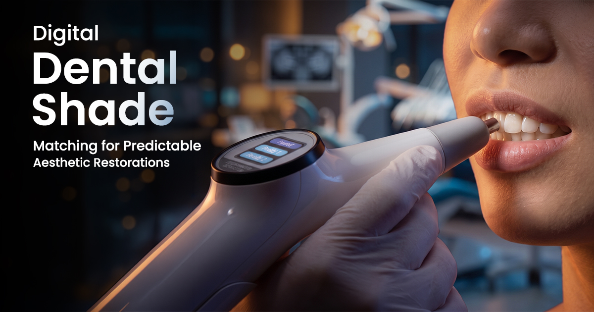
Digital Dental Shade Matching for Predictable Aesthetic Restorations
Precision Beyond the Eye
A dental spectrophotometer is a device that objectively measures tooth color by analyzing reflected light across the visible spectrum, providing precise L*a*b* values. This technology removes the subjectivity of visual shade matching, leading to more predictable and aesthetically superior prosthodontic outcomes.
Table of Contents
How a Dental Spectrophotometer Works
A dental spectrophotometer works by emitting a standardized, full-spectrum white light onto the tooth surface and precisely measuring the wavelength-by-wavelength reflectance of the light that bounces back. Unlike a colorimeter, which measures color based on three broad red, green, and blue filters, a spectrophotometer analyzes the entire visible spectrum (typically 400-700 nm) for a much more detailed and accurate color profile.
This captured spectral data is then converted into coordinates within a three-dimensional color space, most commonly CIELAB (L*, a*, b*). The L* value represents lightness, a* represents the red-green axis, and b* represents the yellow-blue axis. This quantitative data eliminates guesswork and provides the dental laboratory with objective information to create highly accurate prosthodontic products.
- Illumination: Uses a calibrated, stable light source (like an LED) to ensure consistent readings regardless of ambient operatory lighting.
- Spectral Analysis: A diffraction grating or prism inside the device separates the reflected light into its constituent wavelengths, which are then measured by a sensor array.
- Data Output: The device provides not only the closest commercial shade tab equivalent (e.g., VITA 3D-Master or Classical) but also the raw CIELAB values for precise digital communication.
- Shade Mapping: Advanced units can take multiple readings across a single tooth to map the color variations from the cervical third to the incisal edge.
THE DIGITAL SHADE-TAKING PROCESS
The device emits a standardized white light to illuminate the tooth surface consistently.
Light reflects off the tooth, carrying its unique spectral color information.
A sensor measures the reflected spectrum and converts it into objective L*a*b* color data.
Clinical Benefits Over Visual Shade Matching
The primary clinical benefits of digital shade matching are superior accuracy, objectivity, and enhanced communication with the dental laboratory. Visual shade matching with traditional shade guides is notoriously subjective, influenced by factors like operator eye fatigue, color perception deficiencies, and the metameric effect, where colors match under one light source but not another.
Digital instruments bypass these human variables by providing quantitative, repeatable data. This is particularly valuable in the diverse Indian clinical setting, where varying operatory lighting conditions can compromise visual assessments. An objective reading ensures that the shade prescribed is the shade delivered, reducing the rate of remakes and improving patient satisfaction with anterior restorations.
- Eliminates Subjectivity: Readings are independent of the clinician's color vision, experience, or fatigue.
- Overcomes Metamerism: By analyzing the full spectrum, the device prevents color mismatches under different lighting conditions (e.g., daylight vs. indoor light).
- Improves Lab Communication: Sending a digital file with L*a*b* values and a shade map is far more precise than sending a physical shade tab.
- Documentation: Provides a reliable record of pre-treatment shades, which is invaluable for tracking bleaching progress or for medico-legal purposes.
Digital Shade Matching
- Objective and repeatable data
- Unaffected by ambient lighting
- Detailed shade mapping capabilities
- Reduces remakes and saves chair time
- Higher initial investment cost
Visual Shade Matching
- Low cost and simple to use
- No technology learning curve
- Subject to operator variability
- Highly influenced by lighting and fatigue
- Prone to metameric failure
Integrating Digital Shade Taking Into Workflow
To integrate digital shade taking, first ensure the device is fully charged and calibrated according to the manufacturer's instructions, typically against a supplied white reference tile. The patient's tooth should be clean and naturally hydrated; perform the reading before isolation and extensive tooth preparation, as dehydration can temporarily increase the tooth's value (lightness) by 1-2 shade tabs within minutes.
Position the probe tip so it is flush and perpendicular to the flattest area of the tooth's facial surface, usually the middle third. Avoid the incisal edge, where translucency can interfere with the reading. Take at least two or three readings to ensure consistency. For anterior crowns, consider taking separate readings of the cervical, middle, and incisal thirds to create a complete shade map for the technician.
- Step 1: Calibrate: Always perform calibration at the start of the clinical session or before each use.
- Step 2: Prepare Tooth: Ensure the tooth is free of plaque, stain, and lipstick. Keep it moist with saliva.
- Step 3: Position Probe: Place the probe tip flat against the middle third of the tooth, ensuring no gaps that could allow ambient light to leak in.
- Step 4: Capture & Verify: Take the reading and check for consistency. Recapture if readings vary significantly.
- Step 5: Transmit Data: Send the shade file (often a JPEG or proprietary format) along with a standard digital photograph to the lab.
1. Calibrate Device
Before use, calibrate the spectrophotometer against its white standard to ensure baseline accuracy.
2. Select & Prepare Tooth
Choose a clean, hydrated tooth. Take the shade early in the appointment before the tooth dehydrates.
3. Position & Capture
Place the probe tip flush against the middle third of the tooth's facial surface and capture 2-3 readings.
Factors Influencing Digital Shade Reading Accuracy
The most critical factors influencing digital shade reading accuracy are improper calibration, incorrect probe positioning, and the hydration state of the tooth. Failure to calibrate the device before use introduces a systematic error into all subsequent measurements. A well-calibrated instrument provides a reliable baseline for all readings.
Incorrect positioning of the probe is a common source of error. The aperture of the device must be completely flush with the tooth surface to prevent ambient light from contaminating the reading. Any tilt or gap can lead to an inaccurate measurement. Furthermore, tooth dehydration, which begins within minutes of air-drying, significantly alters optical properties by increasing opacity and value (L*), leading to a brighter, less chromatic reading than the tooth's true state.
- Calibration Errors: A dirty or missing calibration tile will lead to skewed results. Calibrate daily.
- Probe Angulation: A probe angled even a few degrees off-perpendicular can cause erroneous readings.
- Tooth Dehydration: Readings on a dehydrated tooth will appear 1-2 shades lighter and less chromatic.
- External Contaminants: Remnants of plaque, calculus, or even lipstick on the tooth surface will be read by the device.
- Ambient Light Leakage: For some non-contact systems or poorly placed contact probes, strong overhead operatory light can interfere.
Avoid Inaccurate Readings
Always take the shade reading before rubber dam placement or prolonged isolation. A tooth that has been air-dried for even 2-3 minutes can become dehydrated, leading to a shade selection that is inaccurately high in value.
Frequently Asked Questions
A dental spectrophotometer provides a more accurate and comprehensive color analysis than a colorimeter. The spectrophotometer measures spectral reflectance across the entire visible light spectrum (400-700 nm), capturing detailed data. In contrast, a colorimeter measures color by filtering light into only three broad data points (red, green, blue), making it more susceptible to errors like metamerism.
Yes, many modern dental spectrophotometers can measure and quantify tooth translucency and opalescence in addition to basic shade. This data is critical for the dental technician when fabricating high-aesthetic restorations like anterior veneers or all-ceramic crowns, as it allows them to replicate the natural optical properties of the adjacent dentition for a seamless integration.
Inconsistent digital shade readings are most often caused by three factors: lack of calibration, variable probe positioning, or changes in tooth hydration. Ensure the device is calibrated before each use, that the probe tip is held perfectly flat and stable against the tooth for every reading, and that the measurements are taken before the tooth has a chance to dehydrate from air exposure.
A dental spectrophotometer should be calibrated according to the manufacturer's guidelines, which typically recommend calibration before each patient use or at the beginning of each day. This simple step, taking less than a minute, ensures that the device's internal sensor is zeroed against a known white standard, compensating for any drift and guaranteeing measurement accuracy.
For full mouth rehabilitation cases where an entirely new shade is being established, the maxillary canine is often used as the reference tooth. Canines typically have the highest chroma (color saturation) and value of all teeth, making them an excellent guide. Taking a digital reading of the untreated canine provides a crucial baseline for the lab technician to build the new aesthetic scheme.
Written by
Dr. Yashasvi Sharma
Contributing Dental Clinician
Dr. Yashasvi Sharma is a contributing Dental professional at Dentalkart Blogs, where she distills chair-side clinical experience into evidence-based, practice-ready guides for Indian dentists. Her work bridges the gap between academic dentistry and everyday practice, translating global research into actionable insights tailored to the realities of Indian clinical settings.
Keep reading
Browse all →
Which Luting Cement for Zirconia Crowns?
Which Luting Cement for Zirconia Crowns?A Clinical Decision Guide The ideal luting cement for zirconia crowns is typically a self-adhesive resin cement, prized

Mastering Direct Chairside Denture Relining: A Clinical
Mastering Direct Chairside Denture Relining: A Clinical ProtocolImprove Fit, Function, and Comfort Direct chairside relining readapts a denture's intaglio surfa
A Clinical Guide to Preventing Dental Fiber Post Debonding
A Clinical Guide to Preventing Dental Fiber Post DebondingTroubleshooting Clinical Failures Fiber post debonding is the most common failure in post and core res
