
Dr. Prerna Sahrawat
Contributing Dental Clinician
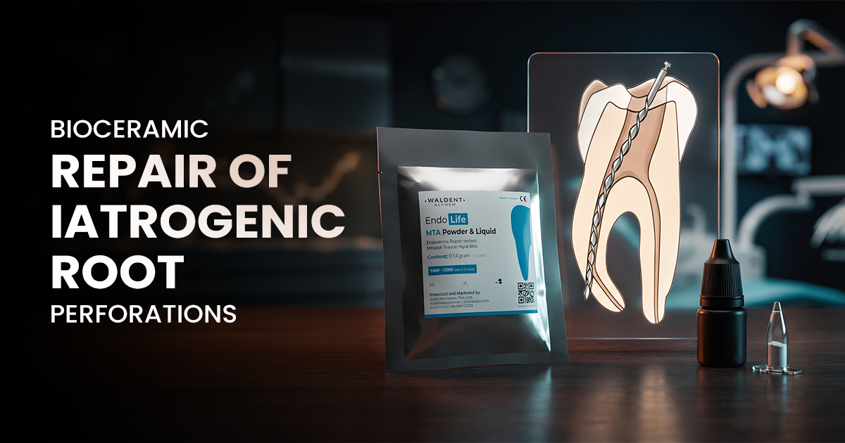
Bioceramic Repair of Iatrogenic Root Perforations
A Modern Endodontic Solution
Bioceramic materials like MTA (mineral trioxide aggregate) and Biodentine offer a highly predictable and biocompatible method for sealing iatrogenic root perforations, which are defects connecting the root canal system to periodontal tissues. Their use is critical for preventing bacterial leakage and salvaging teeth that might otherwise require extraction.
Table of Contents
Diagnosing Strip and Furcal Perforations
- Sudden Hemorrhage: Profuse, uncontrollable bleeding from the canal orifice, which is difficult to stanch with standard hemostatic agents.
- Apex Locator Instability: An apex locator will give a premature, short, or wildly fluctuating reading, often indicating a lateral exit rather than the anatomic apex.
- Direct Vision: With magnification and good lighting (e.g., a dental operating microscope), the perforation site may be directly visible.
- Paper Point Test: A paper point placed in the canal will show blood at a point coronal to the working length, indicating a lateral breach.
Diagnostic Checklist for Root Perforations
Hemorrhage that is difficult to control from within the canal is a primary sign of perforation.
A sudden, short, or unstable reading indicates a breach in the root surface.
Take a periapical X-ray with a file in place to confirm the location of the suspected defect.
A sharp, unexpected pain during instrumentation (if not fully anesthetized) can signal a perforation.
Indications for Bioceramic Repair Materials
- Furcal Perforations: Defects in the bifurcation or trifurcation area of multi-rooted teeth, often caused during access.
- Strip Perforations: Lateral perforations along the canal wall, typically in the 'danger zone' of mandibular molars during instrumentation.
- Apical Perforations: Accidental creation of a foramen coronal to the anatomic apex, often due to over-instrumentation.
- Resorptive Defects: Sealing of internal or external resorptive defects that have perforated into the periodontium.
Bioceramics (MTA/Biodentine)
- Excellent biocompatibility promotes tissue healing.
- Hydrophilic nature allows setting in a moist environment.
- Superior sealing ability prevents bacterial leakage.
- Longer setting time (especially MTA) may require two visits.
- Higher material cost compared to traditional options.
Traditional Materials (Amalgam/IRM)
- Lower material cost and long history of use.
- Fast setting time for single-visit application.
- Potential for corrosion and tissue staining (amalgam).
- Poor seal in the presence of moisture.
- Lack of biocompatibility can impede periodontal healing.
Clinical Protocol for Placing MTA
- Mixing Consistency: MTA should be mixed to a dense, putty-like or wet sand consistency for optimal handling and placement.
- Carrier: Use a specialized carrier like a MAP System, a Dovgan carrier, or the back end of a large paper point to deliver small increments.
- Compaction: Gently compact each increment with a suitable micro-plugger to ensure a dense, void-free seal against the defect walls.
- Moisture: Place a moist cotton pellet over the placed MTA to facilitate its hydrophilic setting reaction; initial setting time is typically 2-4 hours.
Step 1: Hemostasis & Disinfection
Control bleeding with pressure or hemostatic agents. Disinfect the site with 2.5% sodium hypochlorite and rinse thoroughly with saline.
Step 2: Internal Matrix Placement
For larger defects, place an internal resorbable matrix (e.g., collagen sponge) to act as a barrier and prevent MTA over-extrusion.
Step 3: MTA Mixing & Delivery
Mix the MTA powder with sterile water to a wet sand consistency. Deliver small increments to the site using a specialized carrier.
Step 4: Compaction & Sealing
Gently compact the MTA with micro-pluggers. Place a moist cotton pellet over the repair and seal with a temporary restoration for 24 hours.
Assessing Long-Term Repair Success
- Clinical Assessment: Absence of pain, swelling, tenderness to percussion, and periodontal pockets.
- Radiographic Healing: Formation of a continuous lamina dura across the defect and resolution of any pre-existing periradicular radiolucency.
- Functional Stability: The tooth must be in normal function without mobility or discomfort.
- Follow-Up Schedule: Recall appointments should be scheduled at 3, 6, and 12 months, and then annually to monitor the site.
HALLMARKS OF SUCCESSFUL PERFORATION REPAIR
The patient reports no pain, swelling, or discomfort from the treated tooth during function.
Follow-up radiographs show an intact lamina dura and resolution of any previous bone loss.
The tooth is stable, non-mobile, and functions normally within the dental arch.
Frequently Asked Questions
MTA and Biodentine are both calcium-silicate bioceramics, but Biodentine has a much faster setting time of around 12 minutes compared to MTA's 2-4 hours, often allowing for a single-visit procedure. MTA has a longer track record of documented clinical success, while Biodentine offers superior handling characteristics. The choice often depends on the clinical situation and operator preference.
Yes, even large or chronic perforations can often be successfully repaired, provided the associated periodontal lesion is manageable and the tooth is restorable. Success depends on thoroughly debriding the defect site and achieving a complete, void-free seal with an MTA root repair material. A CBCT scan is highly recommended to assess the true extent of the defect before attempting repair.
MTA requires moisture for its setting reaction and typically achieves its initial set in 2 to 4 hours, which is when it can resist displacement. However, it continues to mature and gain compressive strength over several weeks. A moist cotton pellet is placed over the material during the initial setting phase before the final restoration is placed in a subsequent appointment.
The most common reason for failure is bacterial contamination due to an inadequate seal. This can result from poor compaction, leaving voids, or failing to control moisture during placement, which can wash out the material before it sets. Over-extrusion of material into the periodontium can also impair healing and lead to chronic inflammation and failure.
Moisture control is a delicate balance. While bioceramics like MTA are hydrophilic and require moisture to set, excessive bleeding or fluid contamination from the periodontium can wash away the material before it sets. The goal is to stop active hemorrhage, then place the material in a field that is moist but not flooded, often using a moist cotton pellet to provide hydration afterwards.
Written by
Dr. Prerna Sahrawat
Contributing Dental Clinician
Dr. Prerna Sahrawat is a dentist with over 4 years of experience in clinical dentistry and dental content writing. She is passionate about simplifying complex dental concepts into practical, evidence-based content that helps dentists make informed clinical and purchasing decisions while staying updated with the latest advancements in dentistry.
Keep reading
Browse all →
Gauging Canal Status with Absorbent Paper Points
Gauging Canal Status with Absorbent Paper PointsA Clinical Selection Guide Using absorbent paper points for diagnosis provides critical, real-time feedback on c

Ideal Cases for Intraligamentary Anaesthesia
Ideal Cases for Intraligamentary AnaesthesiaAn Evidence-Based Guide Intraligamentary anaesthesia is a supplemental injection technique that forces a local anaes
Biodentine vs. Dycal For Pulp Capping
Biodentine vs. Dycal For Pulp CappingA Case Selection Guide The choice between Biodentine and Dycal as pulp capping materials hinges on bioactivity and seal qua
