
Dr. Yashasvi Sharma
Contributing Dental Clinician
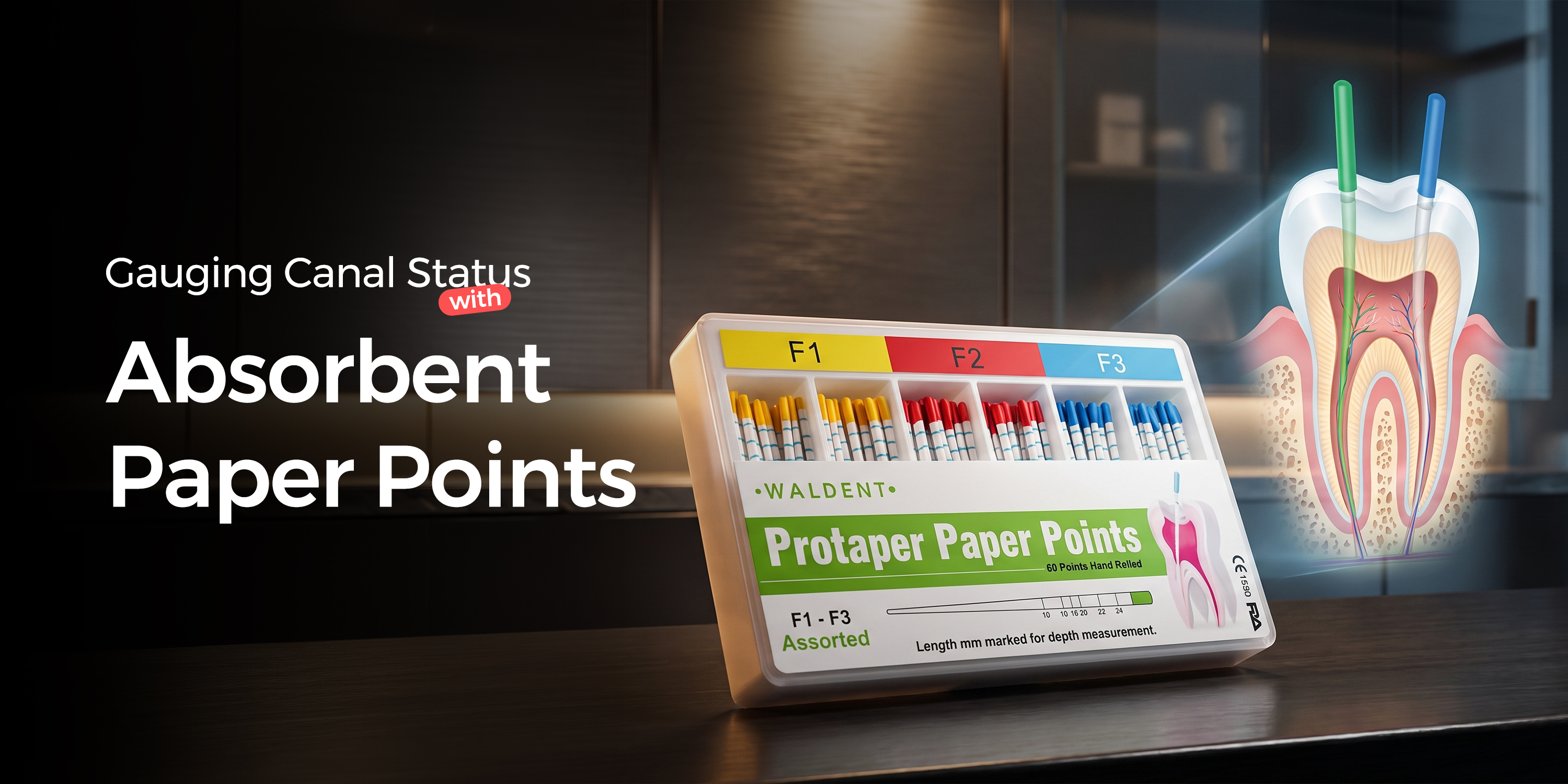
Gauging Canal Status with Absorbent Paper Points
A Clinical Selection Guide
Using absorbent paper points for diagnosis provides critical, real-time feedback on canal moisture and fluid type before obturation. This simple chairside test helps you determine if a canal is truly clean, dry, and ready for a hermetic seal, preventing post-operative failures.
Table of Contents
Defining 'Obturation-Ready' Canal Dryness
A canal is considered sufficiently dry for obturation when a paper point, sized to the master apical file, comes out completely clean and dry after being held at the full working length for 5 seconds. Any moisture, even a hint of dampness on the apical 2-3 mm, indicates that the canal is not yet ready for sealing.
Moisture contamination significantly compromises the physical properties and seal of most common zinc oxide-eugenol and resin-based root canal sealers, creating voids that lead to microleakage and treatment failure. If persistent dampness is observed, especially after thorough irrigation, it's prudent to place an intracanal medicament like calcium hydroxide for 7-14 days rather than proceeding with obturation.
- Apical Focus: The final 2-3 mm of the paper point is the most critical area to inspect for moisture.
- Standardized Timing: Consistently holding the point in place for 5 seconds prevents misleading results from brief contact.
- Serial Confirmation: Always verify dryness with a series of 2-3 consecutive dry points before proceeding to obturation.
- Size Matters: Use a paper point that matches the master apical file diameter to ensure it reaches and effectively dries the apical third.
Chairside Dryness Confirmation
Choose a paper point that corresponds to the master apical file size.
Ensure the point reaches the radiographically or electronically determined working length.
Hold the point passively in the canal for a full 5 seconds.
Carefully examine the apical 2-3 mm under magnification for any sign of moisture or discoloration.
Confirm with at least two subsequent, completely dry paper points before starting obturation.
Differentiating Exudate vs. Haemorrhage
The primary difference between haemorrhage and exudate on a paper point is colour and clinical context. Haemorrhage presents as bright red blood, indicating fresh bleeding, whereas exudate can be serous (clear, straw-coloured), purulent (opaque, whitish-yellow), or sanguineous (blood-tinged fluid).
Haemorrhage is almost always iatrogenic, typically from over-instrumentation with rotary files beyond the apical constriction. This underscores the importance of accurate working length determination with an electronic apex locator, a feature often integrated into modern endomotors. In contrast, exudate signifies persistent inflammation or infection in the periapical tissues, meaning the canal is biologically unprepared for obturation.
- Location of Stain: Blood confined to the very tip of the point strongly suggests instrumentation is 0.5-1 mm too long. Blood smeared along the length may indicate a lateral perforation.
- Colour Clues: Bright red blood means active bleeding. A darker, brownish stain suggests older, resolving haemorrhage.
- Odour as a Sign: A foul or putrescent odour is pathognomonic for purulent exudate from an active anaerobic infection.
| Indicator | Haemorrhage | Exudate |
|---|---|---|
| Appearance | Bright red (fresh) or dark red/brown (old) | Clear, straw-coloured, or opaque yellow/white |
| Source | Iatrogenic (e.g., over-instrumentation, perforation) | Biological (e.g., periapical inflammation, infection) |
| Odour | None | Often present (purulent) best |
| Clinical Action | Re-verify working length, control bleeding | Place intracanal medicament, delay obturation |
What Does a Weeping Canal Indicate?
A weeping canal presents as persistent, non-suppurative fluid flow that continuously wets paper points despite repeated drying efforts. This clear or sero-sanguineous fluid is a periapical exudate, indicating significant inflammation and vascular permeability in the tissues beyond the apex, often associated with a large chronic lesion or a Phoenix abscess.
Attempting to obturate a weeping canal is a common cause of post-operative pain and failure. The constant fluid pressure prevents the sealer from adapting to the canal walls and apex, leading to a poor seal. The standard protocol is to place a calcium hydroxide dressing for 7-14 days to alter the periapical pH, reduce inflammation, and promote the formation of a hard tissue barrier before introducing gutta percha points.
- Source of Fluid: The exudate originates from the periapical tissues, not from within the canal itself.
- Chemical Management: Calcium hydroxide's high pH (approx. 12.5) helps denature inflammatory proteins and has an antibacterial effect.
- Mechanical Blockage: Before placing medicament, confirm apical patency to ensure a physical blockage of debris is not trapping the fluid.
- Systemic Considerations: In rare, stubborn cases, consider if systemic patient factors, like uncontrolled diabetes, are delaying periapical healing.
Managing Paper Point Findings
Recognize the Limits of Paper Point Diagnosis
Do not rely on a dry paper point as the sole indicator of canal sterility, especially in cases with complex anatomy, suspected fractures, or long-standing chronic infections. The paper point only samples the planktonic bacteria and fluid in the main canal lumen; it provides no information about the adherent bacterial biofilms within dentinal tubules, lateral canals, or isthmuses.
A dry point is a prerequisite for obturation, but it is not a guarantee of disinfection. True canal sterilization relies on the chemical action of irrigants, the mechanical removal of tissue by instrumentation, and proper sealing with three-dimensional obturation systems. A paper point is a test for dryness, not a test for sterility.
- Biofilms: A canal can be perfectly dry to a paper point yet still harbor a significant, mature biofilm on its walls.
- Vertical Root Fracture (VRF): While a VRF can sometimes cause persistent weeping, the paper point finding is non-specific. Diagnosis requires correlation with probing depths, radiographic evidence (J-shaped lesion), and direct visualization.
- Anatomical Complexity: In C-shaped canals, fins, or webs, the paper point will only contact a small fraction of the total surface area, giving a potentially misleading impression of the canal's overall status.
- Microleakage: A paper point cannot detect coronal microleakage from a faulty temporary restoration, which can re-infect the canal between appointments.
Clinical Misinterpretations to Avoid
Never assume a dry paper point equals a sterile canal. It only indicates the absence of free-flowing fluid at a single moment. Disinfection is achieved through a multi-step protocol of chemo-mechanical preparation, not by achieving a dry reading on a paper point.
Conclusion: Reading the Point Correctly
The single most important judgement is that an absorbent paper point is an indispensable chairside tool for assessing moisture, but it is not a substitute for comprehensive endodontic diagnosis. Its findings must be correlated with radiographic evidence, clinical signs, and a thorough understanding of canal anatomy to make the final, critical decision to obturate. A misread point can be the difference between long-term success and predictable failure.
- Dryness Standard: A canal is ready only when a paper point held for 5 seconds to the full working length emerges completely clean and dry.
- Weeping Canal Rule: Never obturate a weeping canal. The correct protocol is to place a calcium hydroxide dressing for a minimum of 7-14 days and then reassess.
- Bleeding Indicator: Blood isolated to the apical tip suggests instrumentation is likely 0.5-1.0 mm beyond the apical constriction; re-verify and adjust your working length.
- Diagnostic Limit: A dry point does not confirm sterility in complex anatomy like C-shaped canals or isthmuses, where biofilms persist.
Frequently Asked Questions
Absorbent paper points are primarily used to dry the root canal system after cleaning and shaping, just before obturation. Their critical secondary function is diagnostic: they help assess the canal for haemorrhage or persistent exudate, which indicates whether the canal is biologically ready to be sealed.
Pus (purulent exudate) will appear opaque, thick, and typically yellowish-white on a paper point, often with a distinct foul odour from anaerobic bacteria. In contrast, serous fluid is clear, watery, and straw-coloured, indicating periapical inflammation without the same level of active, suppurative infection.
There is no fixed number; use as many as it takes. Introduce a series of appropriately sized absorbent paper points one by one until one comes out completely dry after being held at the working length for 5 seconds. This usually takes 3-5 points for a well-prepared canal, but a weeping canal may require many more before you decide to place a medicament instead.
The ideal paper point should match the size of the master apical file (MAF). For example, if you finished shaping with a size 35/0.04 file, use a size 35 paper point. A point that is too small won't reach the apex effectively, while one that is too large will bind coronally and fail to dry the critical apical third.
The most common mistake is failing to place the paper point to the full, verified working length. If the point is short, it can give a false negative for dryness, leaving moisture in the apical 2-3 mm. This residual moisture can compromise the seal of endodontic products like sealers and ultimately lead to treatment failure.
Written by
Dr. Yashasvi Sharma
Contributing Dental Clinician
Dr. Yashasvi Sharma is a contributing Dental professional at Dentalkart Blogs, where she distills chair-side clinical experience into evidence-based, practice-ready guides for Indian dentists. Her work bridges the gap between academic dentistry and everyday practice, translating global research into actionable insights tailored to the realities of Indian clinical settings.
Keep reading
Browse all →
Ideal Cases for Intraligamentary Anaesthesia
Ideal Cases for Intraligamentary AnaesthesiaAn Evidence-Based Guide Intraligamentary anaesthesia is a supplemental injection technique that forces a local anaes

Biodentine vs. Dycal For Pulp Capping
Biodentine vs. Dycal For Pulp CappingA Case Selection Guide The choice between Biodentine and Dycal as pulp capping materials hinges on bioactivity and seal qua

Case Selection for Electric Pulp Testing
Case Selection for Electric Pulp TestingA Diagnostic Decision Guide An electric pulp tester (EPT) assesses pulp nerve response using a graduated electrical curr