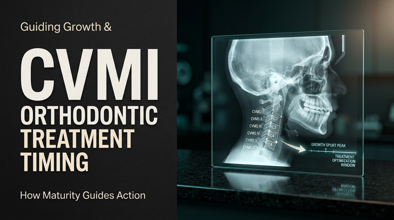
How CVMI in Orthodontics Helps you time Growth Modification

Dr. Gurjeet
Contributing Dental Clinician
What is CVMI in Orthodontics and How Do You Read the Six Stages?
Cervical vertebral maturation (CVM) guides timing by staging a patient from CS1 to CS6 using the shape of C2, C3 and C4 on the lateral cephalogram already taken for diagnosis. The pubertal mandibular growth peak falls between CS3 and CS4. Growth modification for mandibular deficiency is generally most effective when treatment spans that interval; by CS5 and CS6 the opportunity has largely passed.
That makes CVM a timing tool, not a treatment selector. It answers when, not what.
CVM or CVMI? Getting the Terms Right
Clinicians, and search engines, use the two terms interchangeably, but they describe two different staging systems:
CVMI (cervical vertebral maturation index) usually refers to the six-stage index described by Hassel and Farman (1995): initiation, acceleration, transition, deceleration, maturation and completion.
CVM in current practice usually means the revised method of Baccetti, Franchi and McNamara (2005), with stages CS1 to CS6.
Both read the same vertebrae and both have six stages, but the stage boundaries are not identical, so a "stage 3" in one is not automatically a "CS3" in the other. This article describes the Baccetti CVM method throughout. Whichever system your clinic uses, name it in the notes so that colleagues read the stage the same way.
Why Chronological Age Misleads
Two 11-year-olds in the same class can be two years apart in skeletal maturity. Girls typically reach each maturational stage earlier than boys, and variation within each sex is wide. A functional appliance started by age alone may be delivered too early, wearing out the child's cooperation before the spurt arrives, or too late, after the mandibular growth it depends on has slowed.
Biological indicators correct for this. Hand-wrist radiographs were the traditional option, but they need an extra exposure. CVM uses the lateral cephalogram that is already part of the orthodontic record, which is why it has become the routine choice. A hand-wrist film is now reserved for the occasional case where the CVM stage remains genuinely unclear and the treatment decision depends on it.
The Six Stages and What Each Means for Timing
The Baccetti method assesses two features: whether there is a concavity at the lower border of C2, C3 and C4, and the shape of the C3 and C4 bodies.
Stage | Radiographic criteria | Relation to the growth peak |
CS1 | Lower borders of C2, C3, C4 flat; C3 and C4 trapezoid | Peak expected no earlier than about 2 years later |
CS2 | Concavity at C2 only; C3 and C4 still trapezoid | Peak expected about 1 year later |
CS3 | Concavities at C2 and C3; bodies trapezoid or horizontal rectangular | Peak expected within about 1 year |
CS4 | Concavities at C2, C3 and C4; both bodies horizontal rectangular | Peak occurred within the previous 1 to 2 years |
CS5 | At least one of C3 or C4 square | Peak ended at least 1 year earlier |
CS6 | At least one of C3 or C4 vertical rectangular | Peak ended at least 2 years earlier |
Intervals as described by Baccetti, Franchi and McNamara (2005). They are population averages, not a calendar for an individual patient.

A common misreading places CS4 "at the peak". In the original description the peak lies between CS3 and CS4; CS4 means it has recently passed. For a functional appliance case, that distinction can decide whether treatment starts now or the window is already narrowing.
Reading the Stage Reliably
Start with the film
C2 to C4 must be clearly visible and sharp, and C4 must not be cut off by the collimator or hidden by a thyroid collar. Head rotation creates double contours on the lower borders, which can mimic or hide concavities. If you cannot see the lower border of C4 clearly, you cannot separate CS3 from CS4, so do not guess. Good positioning on the cephalometric X-ray unit matters here as much as it does for the skeletal tracing.
Then work in a fixed order
Inspect the lower borders of C2, C3 and C4 for a concavity, working downwards from C2.
Classify the C3 and C4 body shapes: trapezoid, horizontal rectangle, square or vertical rectangle.
Assign the stage from the criteria table, not from a general impression.
Cross-check against age, sex, recent height gain and secondary sexual characteristics where these are known.
The cross-check is where most errors are caught. A 9-year-old boy staged at CS5, or a 15-year-old girl staged at CS1, should send you back to the film before the stage is trusted.
Expect observer variability
CVM staging is less reproducible than its tidy table suggests, and the literature is genuinely divided. Gabriel and colleagues (2009) titled their study "Cervical vertebrae maturation method: poor reproducibility", after finding low agreement among trained orthodontists when common methodological shortcuts were removed. Other studies report better figures: Rainey and colleagues (2016) found substantial agreement after structured training, and a 2024 meta-analysis reported satisfactory overall reliability, with higher agreement in studies where the vertebrae were traced rather than staged by eye.
The practical message is the same either way. The CS2 to CS4 range, exactly where timing decisions are made, is the hardest to call. Stage from the original digital image rather than a compressed print, trace the vertebrae if a decision hangs on the stage, have a second clinician stage borderline films independently, and prefer serial staging to a single reading when you can.

Worked Timing Scenarios
Hypothetical patients, both Class II with mandibular retrognathism on cephalometric analysis.
Patient A | Patient B | |
Age and sex | 11 years, female | 11 years, male |
CVM stage | CS3 | CS1 |
Expected peak | Within about 1 year | No earlier than about 2 years |
Timing implication | Window is open; a growth-modification phase started now can span the peak | A functional appliance started now may run well before the peak; consider monitoring or addressing other problems first |
Restage | If treatment is deferred, recheck at the next records | Recheck at the next indicated cephalogram |

Same age, same malocclusion, different timing. If a functional appliance phase is expected to last around 12 months (a hypothetical planning figure), Patient A's treatment would likely include the peak, while Patient B's would likely finish before it begins.
For Class III cases the logic shifts earlier. Maxillary protraction with a facemask is generally reported to produce more skeletal effect when started early, typically in the prepubertal stages (CS1 to CS2), before the circummaxillary sutures mature.
What CVM Cannot Tell You
CVM estimates how much growth remains. It does not show the direction of growth, which jaw is at fault, the vertical pattern, crowding or tooth size. A Class II at CS3 points to different options from the same Class II at CS6, but only cephalometry, model analysis and Angle's classification identify what the problem actually is.
It also does not guarantee a response. Growth modification outcomes vary between patients and appliance types, and the size of the skeletal change with functional appliances is still debated. CVM improves the odds that treatment coincides with available growth; it does not promise how much of that growth will be redirected.
Documenting the Stage
Record the CVM stage, the method used, the date of the cephalogram, any film-quality limitation (for example, "C4 lower border partially obscured"), age and sex, and the planned restaging point. An undated CVM stage is close to useless, because the stage only means something relative to when the film was taken. Store the image alongside dated clinical photographs from an intraoral camera and digital models or scans, so the growth phase and the occlusion can be compared at each review.
Frequently Asked Questions
Only when a pending decision depends on it, such as whether to start a functional appliance phase now or wait. Under the ALARA principle, a repeat radiograph needs a clinical justification. Height records and clinical progress can often support the decision in the meantime.
It helps confirm that growth has largely finished, which matters most in Class III cases, where late mandibular growth can undermine a surgical result. CS6 suggests the peak ended at least two years earlier, but surgical timing usually also relies on serial cephalograms showing stability, and on age, rather than on a single CVM reading.
Yes, and it is still informative. Even without growth modification, knowing whether a patient is before or after the peak helps predict sagittal and vertical changes during treatment, which affects anchorage, Class II elastic planning and the expected stability of the final occlusion.
Written by
Dr. Gurjeet
Contributing Dental Clinician
Dr. Gurjeet is a practising dental clinician contributing to Dentalkart Blogs, sharing chair-side experience, treatment protocols, and equipment insights for the wider dental community.
Keep reading
Browse all →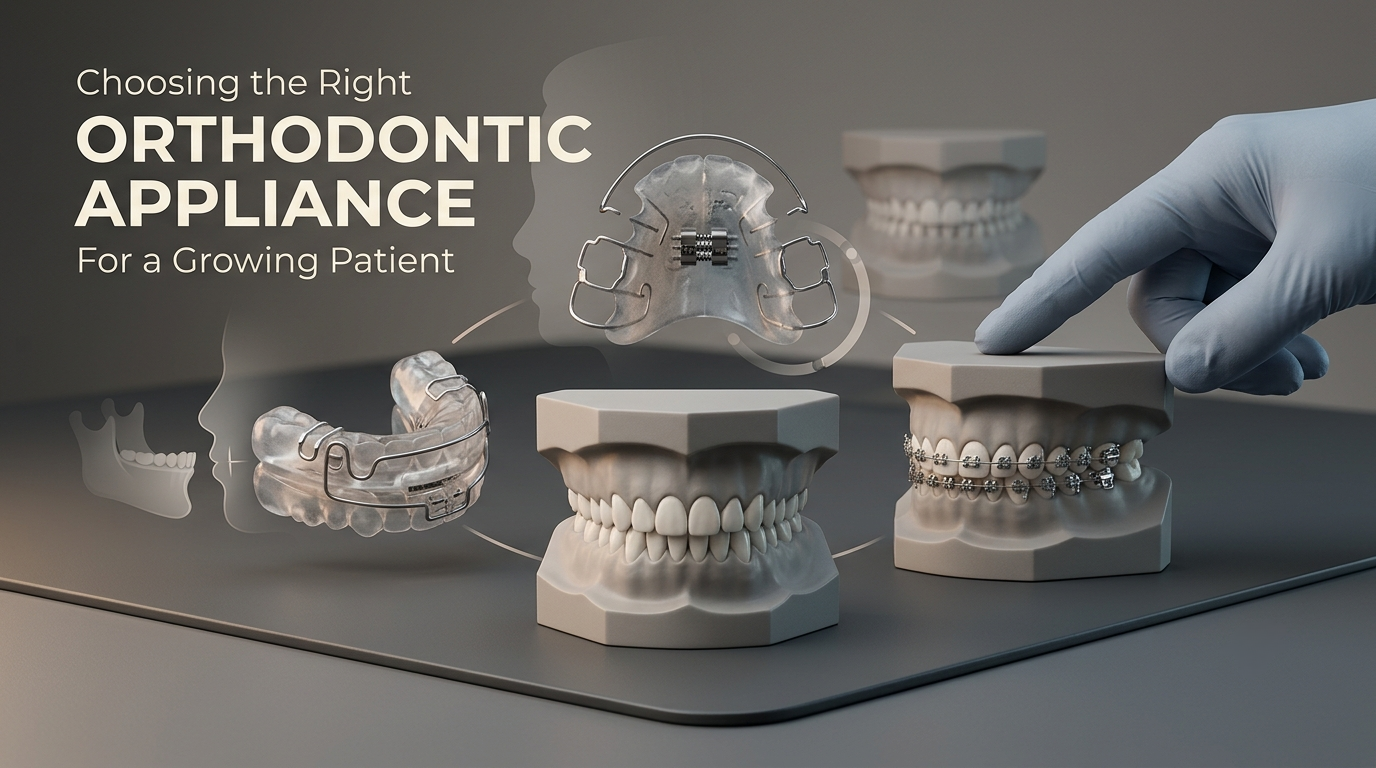
How to Choose Orthodontic Appliances for Growing Patients
How Do You Choose Between a Functional, Removable, or Fixed Orthodontic Appliance?In a growing patient, choose the appliance by the level of the problem first,
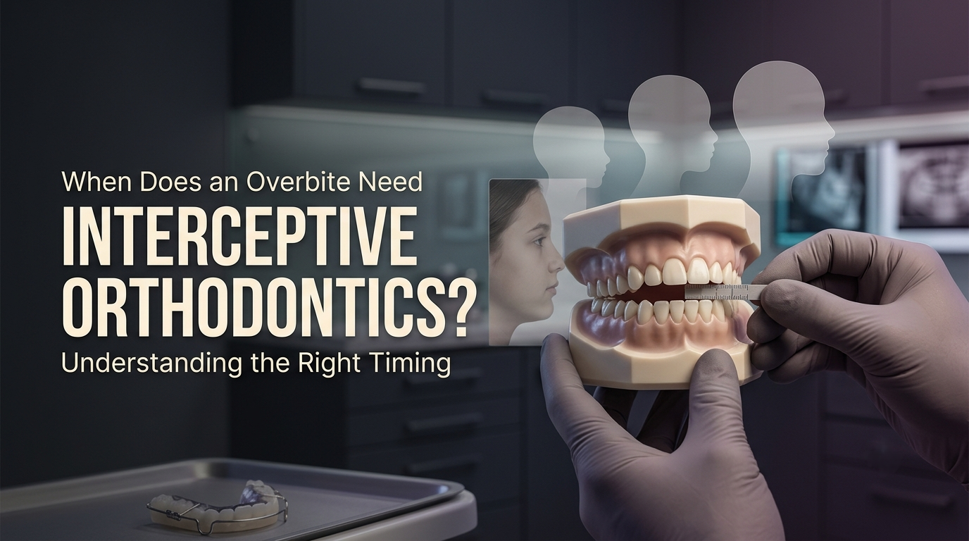
When Interceptive Orthodontics Is Needed for an Overbite
When Does an Overbite Case Call for Interceptive Orthodontics Instead of Waiting?Interceptive orthodontics is justified in an overbite case in two situations: w
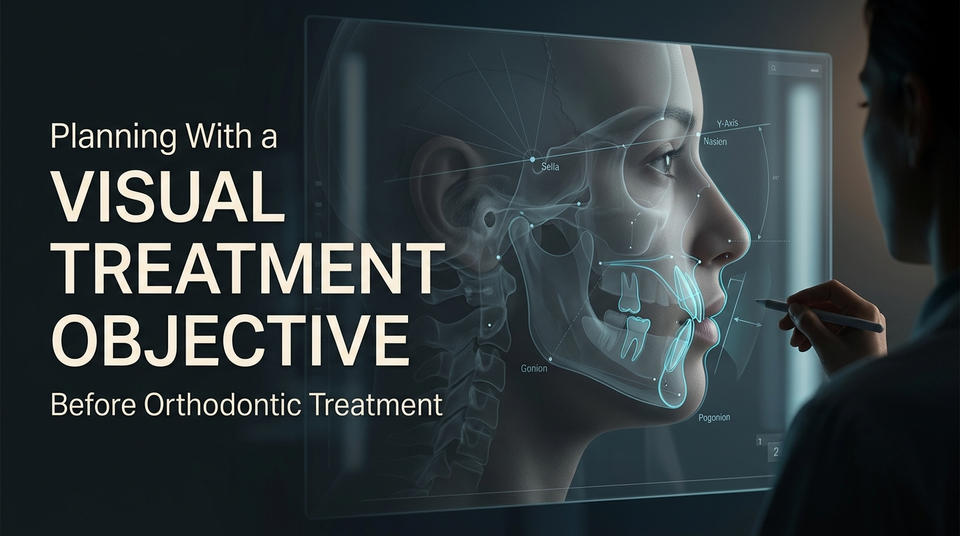
How VTO in Orthodontics Helps Plan Treatment and Outcomes
How Is a Visual Treatment Objective (VTO) Used to Plan an Orthodontic Case?A Visual Treatment Objective is used to test a plan before treatment starts. On the l