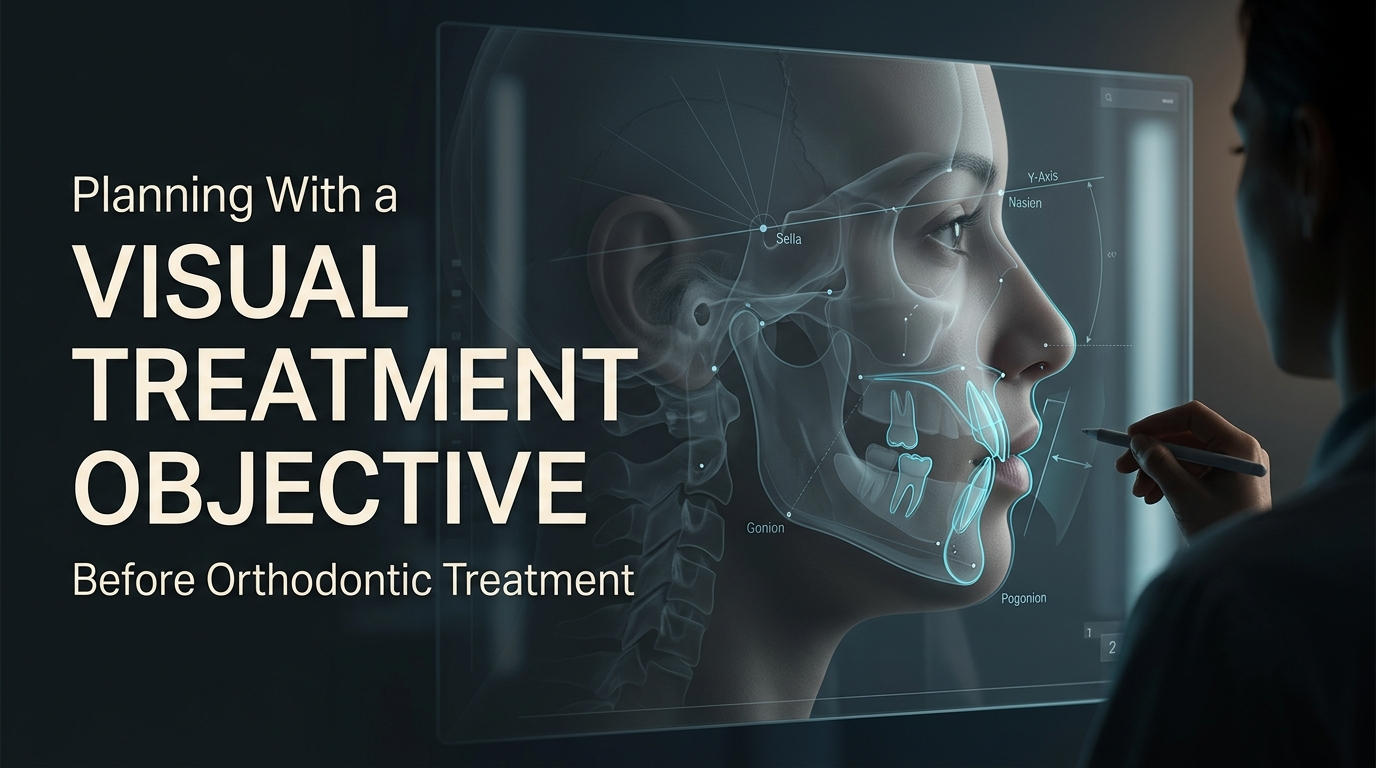
How VTO in Orthodontics Helps Plan Treatment and Outcomes

Dr. Gurjeet
Contributing Dental Clinician
How Is a Visual Treatment Objective (VTO) Used to Plan an Orthodontic Case?
A Visual Treatment Objective is used to test a plan before treatment starts. On the lateral cephalometric tracing, you forecast growth over the treatment period, set the lower incisor position, then place the molars, upper incisors and soft tissue for the intended result. The VTO shows how much of the correction must come from growth, tooth movement or surgery, and whether that combination is realistic.
If the plan depends on more growth than the patient has left, or on moving teeth beyond what the alveolar bone allows, the VTO shows it on paper rather than eighteen months into treatment.
What a VTO Actually Adds Up
Ricketts popularised the VTO as a way to make the treatment goal visible. It is not a single measurement. It is the sum of three contributions:
In other words, the final position is the current position, plus expected growth, plus planned tooth movement, plus any surgical movement.
Every millimetre of correction has to come from one of those sources. The value of the VTO is that it forces you to assign each millimetre explicitly.
Growth forecast and the Y-axis
The Y-axis runs from sella to gnathion. Its angle to Frankfort horizontal, described by Downs, averages about 59° with a range of roughly 53° to 66°. A smaller angle suggests a more horizontal growth direction, with the chin coming forward; a larger angle suggests vertical growth, with the chin moving more downward than forward.
That matters for Class II correction. In a patient with a Y-axis near 66°, mandibular growth contributes less to reducing overjet, because more of it is expressed vertically. A VTO built on an average growth assumption for that patient will overstate what growth can deliver.
Growth only counts if it is still available, so anchor the forecast to skeletal maturity (see our article on CVM staging), not to age.
The envelope of discrepancy
Proffit and Ackerman's envelope of discrepancy describes how far teeth can be moved by orthodontics alone, how much further growth modification extends that in a growing patient, and the larger range orthognathic surgery allows. For anteroposterior incisor movement by orthodontics alone, the figure in Proffit's Contemporary Orthodontics gives approximately:
Arch | Retraction | Advancement |
Maxillary incisors | About 7 mm | About 2 mm |
Mandibular incisors | About 2 mm | About 3 mm |
The two arches are limited differently. Upper incisors can be retracted a long way into extraction space but advanced only a little before lip support and labial bone become a problem. Lower incisors are limited in both directions by the narrow symphysis and thin labial and lingual cortical plates, which is why the mandibular arch is often called the limiting arch. These are guideline figures, not guarantees; in a patient with a thin symphysis or thin gingiva, the safe range is smaller.

The VTO's job is to check whether the planned tooth positions fall inside the relevant envelope for each arch.
Building the VTO, Step by Step

Start from a sound tracing. Correct head position, clear landmarks, and the growth stage recorded alongside.
Forecast growth for the treatment period. Use the Y-axis direction and maturity stage. For an adult, growth is effectively zero.
Set the lower incisor first. Its position governs arch length, lip support and long-term stability, and its envelope is the tightest. Ricketts used the lower incisor's relationship to the A-Pog line (about +1 mm ± 2 mm) as a reference.
Place the molars and upper incisors for a Class I relationship with target overjet and overbite of about 2 to 3 mm, and an interincisal angle in the region of 130°.
Draw the soft tissue. Predict lip and chin change from the planned incisor and skeletal movement, accepting that soft-tissue response varies between patients.
Test against the envelope. Is each planned movement achievable by the intended method, in each arch?
Worked Example: Where Does the Overjet Correction Come From?
Hypothetical cases. Growth contribution is an assumption, not a prediction.
Case A: growing patient, Class II Division 1, overjet 9 mm, target 2.5 mm. Correction needed: 6.5 mm.
Source | Contribution |
Differential mandibular growth (assumed; favourable Y-axis, pre-peak) | 2.5 mm |
Upper incisor retraction | 3.0 mm |
Lower incisor advancement | 1.0 mm |
Total | 6.5 mm |
Upper retraction of 3 mm (envelope about 7 mm) and lower advancement of 1 mm (envelope about 3 mm) both sit comfortably inside the orthodontic envelope. The plan is feasible, but it depends on that 2.5 mm of growth. If growth disappoints, the shortfall falls on the upper incisors.
Case B: same malocclusion, adult. Correction needed: 6.5 mm; growth 0 mm.
Source | Contribution |
Upper incisor retraction | 5.5 mm |
Lower incisor advancement | 1.0 mm |
Total | 6.5 mm |
Still inside the envelope, but 5.5 mm of retraction usually needs extraction space and careful anchorage, and the VTO must show whether the resulting lip and nose relationship suits the face.

Case C: adult, overjet 12 mm, target 2.5 mm. Correction needed: 9.5 mm; growth 0 mm.
Source | Contribution | Orthodontic envelope |
Upper incisor retraction | 7.0 mm | About 7 mm (fully used) |
Lower incisor advancement | 2.5 mm | About 3 mm (almost fully used) |
Total | 9.5 mm | About 10 mm available |
On paper, the correction just fits, with only about half a millimetre to spare. In practice, that means taking the upper incisors to the very limit of retraction and the lower incisors almost to the limit of advancement, at the same time, in a patient with no growth to help. Both movements at their extremes carry real risk to labial bone, gingival support and root resorption, and to the profile: large upper retraction can flatten the lips, and lower proclination of this size is often unstable. A VTO that only works when both arches are pushed to the edge of the envelope is the signal to discuss surgical correction with the patient alongside camouflage, not a green light for camouflage.
Reference Values Used in a VTO
Feature | Common reference | Role in the VTO |
Lower incisor to A-Pog | About +1 mm ± 2 mm (Ricketts) | Sets arch length and lower lip support |
Interincisal angle | About 130° | Influences bite depth and stability |
Y-axis to FH | About 59° (range about 53° to 66°) (Downs) | Indicates growth direction |
Overjet and overbite | About 2 to 3 mm each | Defines the finished occlusion |
Lip position | Balanced for the individual face | Cross-check with facial analysis |
These are guides for a balanced finish, not targets to chase. A VTO that meets every norm but places the lower incisors outside the symphysis, or flattens an already straight profile, is a worse plan than one that accepts a deviation.
What a VTO Changes in the Plan
The VTO turns the treatment choice into a question of quantity. If tooth movement alone covers the correction comfortably within the envelope, treatment can proceed without relying on growth. If it needs growth, the CVM stage decides whether the window is open and when to start. If it exceeds what growth and tooth movement together can safely provide, or only fits at the edge of the envelope, surgical planning becomes the realistic route, with incisor decompensation built into the pre-surgical phase.
It also shapes the mechanics. A VTO assumes the teeth reach their goal positions; the bracket prescription and archwire sequence must actually deliver them. One detail worth knowing is the "wagon wheel" effect described by Rauch: when torque is expressed on anterior teeth in a rectangular wire, it also changes their mesiodistal angulation (tip). Planned incisor torque may therefore need compensating tip in the prescription or wire to keep the incisors where the VTO put them.
Where VTOs Go Wrong
The most common errors are overestimating growth, ignoring growth direction, chasing cephalometric norms instead of the face, and treating the VTO as a promise to the patient. Growth varies between individuals, and soft-tissue response to incisor movement varies even more. Present the VTO to patients as a plan and a range, not a guaranteed profile.
Revisit the VTO, too. A progress lateral cephalogram, taken when a decision depends on it, lets you superimpose the actual change on the planned change and adjust mechanics before the gap widens.
Frequently Asked Questions
Digital cephalometric software builds VTOs faster and makes superimposition easier, but its soft-tissue predictions rely on average ratios between hard- and soft-tissue movement. Studies of prediction programs generally show acceptable accuracy for hard-tissue positions and wider error for the lips and nasal tip. Treat predicted profiles as illustrations, especially when you show them to patients.
No. An aligner set-up, built from an intraoral scan on digital dentistry platforms, simulates tooth movement within the arches. It does not model skeletal growth, the bony envelope or soft-tissue change on the profile. For Class II or Class III cases, a cephalometric VTO is still needed to decide whether tooth movement alone is a sensible plan.
Yes, and that is often where it is most useful. With growth set to zero, every millimetre of correction must come from tooth movement, so the VTO shows directly whether the planned incisor positions stay within bone and whether the resulting lip position is acceptable.
Written by
Dr. Gurjeet
Contributing Dental Clinician
Dr. Gurjeet is a practising dental clinician contributing to Dentalkart Blogs, sharing chair-side experience, treatment protocols, and equipment insights for the wider dental community.
Keep reading
Browse all →
How to Choose Orthodontic Appliances for Growing Patients
How Do You Choose Between a Functional, Removable, or Fixed Orthodontic Appliance?In a growing patient, choose the appliance by the level of the problem first,
When Interceptive Orthodontics Is Needed for an Overbite
When Does an Overbite Case Call for Interceptive Orthodontics Instead of Waiting?Interceptive orthodontics is justified in an overbite case in two situations: w
Facial Divergence in Orthodontics and Treatment Planning
How Does Facial and Photographic Analysis Change an Orthodontic Treatment Plan?Facial analysis changes the plan by setting soft-tissue limits on tooth movement.