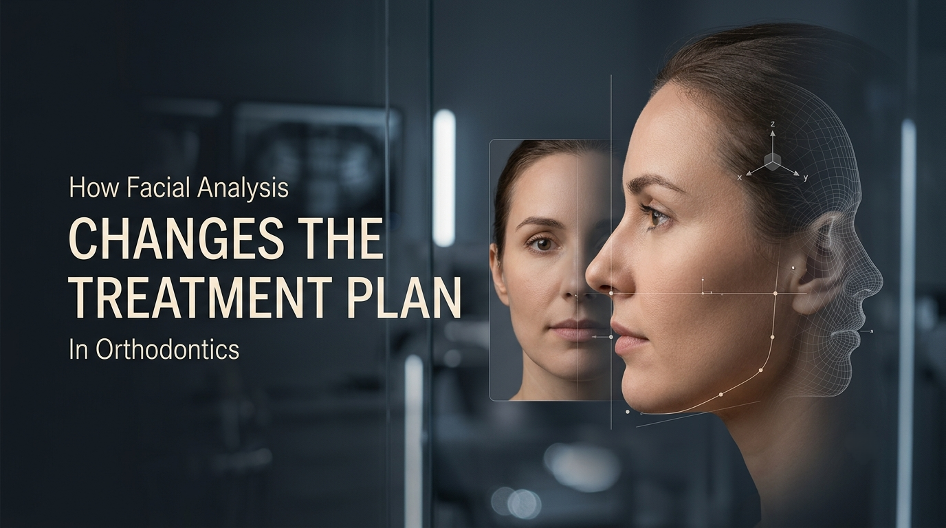
Facial Divergence in Orthodontics and Treatment Planning

Dr. Gurjeet
Contributing Dental Clinician
How Does Facial and Photographic Analysis Change an Orthodontic Treatment Plan?
Facial analysis changes the plan by setting soft-tissue limits on tooth movement. Facial divergence, profile convexity, lip support, facial thirds, incisor display and symmetry decide whether incisors can be retracted or proclined, whether extraction or non-extraction suits the face, whether vertical control is needed, and whether growth modification or surgery is the more appropriate route. Identical cephalometric values can therefore lead to different plans in different faces.
Radiographs describe the skeleton. Patients judge the result by their face. A plan that ignores the face can be technically correct and still disappoint.
What the Face Tells You That the Ceph Does Not
A lateral cephalogram measures skeletal relationships and incisor inclination. It does not show how the lips drape over the teeth at rest, how much tooth shows on smiling, or whether a chin deviation is visible from the front. Facial analysis covers three planes:
Sagittal: facial divergence, profile convexity, lip position, nose and chin prominence.
Vertical: facial thirds, lower-face proportions, lip competence, incisor display at rest and on smiling.
Transverse: symmetry, midlines, chin position and smile width.
What Facial Divergence Means
Facial divergence is a specific term, and it is often confused with profile convexity. It was described by Hellman and is used in Proffit's facial profile analysis. It describes the forward or backward inclination of the lower face relative to the forehead, judged in profile with the patient in natural head position:
Anterior divergent: the lower face, and the chin in particular, inclines forward relative to the forehead.
Straight (orthognathic): the lower face sits roughly in line with the forehead.
Posterior divergent: the lower face, and the chin, inclines backward relative to the forehead.
A simple way to see it chairside: imagine a line from soft-tissue glabella (the forehead) to soft-tissue pogonion (the chin) and look at how it tilts against true vertical.
Divergence is not the same as convexity. Convexity compares the three points of forehead, base of the nose and chin, and tells you whether the profile is straight, convex or concave. Divergence tells you which way the whole lower face leans. A patient can have a straight profile that is posteriorly divergent, or a convex profile that is anteriorly divergent. Divergence also varies naturally between ethnic groups, so it is not abnormal in itself. It matters because it changes how a given incisor or chin position will look, and how any skeletal change will read on the face. For instance, a posteriorly divergent face with a retrusive chin often reflects a mandibular problem, whereas an anteriorly divergent face with the same overjet may point more to the maxilla or incisors.

Taking Photographs You Can Actually Compare
Analysis is only as reliable as the photographs. Three rules matter more than which camera you buy.
Use natural head position (NHP). Ask the patient to look at a distant point, or into their own eyes in a mirror at eye level, with the head relaxed. Frankfort horizontal is a poor substitute in many patients because porion and orbitale positions vary.
Control head rotation. Pitch (nodding) changes profile convexity, divergence and lip position. Roll (tilting) creates false vertical asymmetry. Yaw (turning) distorts the profile and makes one side of the face look fuller.
Keep the geometry constant. Use the same camera-to-subject distance, focal length and lighting at every visit. A medium telephoto macro lens (around 100 mm on a full-frame camera) is commonly used for facial photographs because it reduces perspective distortion. If you intend to measure in millimetres, include a scale in the frame or calibrate to a known dimension. Uncalibrated photographs support ratios and angles, not absolute distances.
The standard set is frontal at rest, frontal smiling, right profile at rest, an oblique smiling view, and intraoral views captured with retractors, mirrors and a good intraoral camera or DSLR setup. Lips should be relaxed and teeth lightly together for the rest views; strained lip closure hides incompetence and alters profile readings.
Reference Values Worth Measuring
Feature | Common reference | Source or note |
Facial thirds | Upper, middle and lower thirds roughly equal | Classical proportion; varies with age and ethnicity |
Lower third split | Subnasale to stomion about one-third; stomion to soft-tissue menton about two-thirds | Widely used clinical proportion (Proffit and colleagues) |
Facial convexity (G-Sn-Pg′) | 12° ± 4° | Legan and Burstone (1980) |
Nasolabial angle | About 90° to 110° | Commonly cited range (Proffit and colleagues); Legan and Burstone reported about 102° ± 8°; influenced by nasal tip shape |
Upper incisor display at rest | About 2 to 4 mm | Decreases with age as the upper lip lengthens |
Lip competence | Lips meet at rest without mentalis strain | A small interlabial gap can be normal in adolescents |
These are population references, not targets. They differ between ethnic groups, between sexes and with age. A value outside the range is a reason to look closer and to correlate with the ceph and the patient's own concerns, not a diagnosis on its own.
Worked Example: Reading the Vertical Proportions
Hypothetical measurements from a calibrated frontal photograph.
Measurement | Value |
Upper third (trichion to glabella) | 60 mm |
Middle third (glabella to subnasale) | 62 mm |
Lower third (subnasale to soft-tissue menton) | 74 mm |
Total face height | 196 mm |
Lower third share | About 38% (equal thirds would be about 33%) |
Subnasale to stomion | 22 mm |
Upper lip share of lower third | About 30% (reference about 33%) |

The lower third is about a fifth longer than the other two. Within it, the upper lip segment is proportionally short and the lip-and-chin segment carries the excess. That raises three questions: is there vertical maxillary excess, increased lower anterior face height on the ceph, or simply a short upper lip? Each points to different mechanics. If the cephalometric vertical pattern confirms a long lower face, molar extrusion becomes a risk to avoid, because mechanics that extrude posterior teeth would worsen exactly what the patient sees in the mirror.
A second example, in profile: a soft-tissue convexity of 22° sits 10° above the Legan and Burstone mean, or 2.5 standard deviations. That is a strong flag to check whether the convexity comes from a retrusive mandible, a prominent maxilla or proclined incisors pushing the lips forward, and the patient's facial divergence helps answer that.
How Facial Findings Change the Plan

Incisor position and extraction decisions
The lips rest on the incisors, so moving the incisors changes lip support. Retracting upper incisors in a patient with an already flat profile, an obtuse nasolabial angle and thin lips can age the face. In a convex profile with lip strain and procumbent lips, retraction may improve lip competence. That is why the extraction decision cannot be made from crowding and cephalometric values alone. The lip response to incisor retraction varies widely with lip thickness and tone, so keep predictions conservative.
Growth modification
A convex, posteriorly divergent profile with a retrusive chin in a growing patient suggests a mandibular deficiency that may respond to growth modification, if the growth stage supports it. The same profile in an adult points towards camouflage or surgery.
Vertical control
A long lower third with lip incompetence and excessive incisor display on smiling argues for mechanics that control molar extrusion. A short lower third with a deep labiomental fold argues the opposite.
Surgical referral
Marked asymmetry, severe convexity or concavity, or a discrepancy where camouflage would worsen the face rather than improve it, belongs in a surgical-orthodontic discussion.
Mistakes That Distort the Reading
The common errors are technical rather than interpretive: head tilted or turned, lips strained, distance changed between visits, and millimetres measured from uncalibrated images. Interpretive errors follow: treating references as goals, applying norms from one population to another, confusing convexity with divergence, and assuming every chin deviation is skeletal. A mandibular shift on closure can produce a facial asymmetry that disappears in centric relation, so check the bite before calling it skeletal.
Read facial findings alongside the lateral cephalogram, study models or digital scans, Angle's classification and the growth stage, and combine everything into one problem list before choosing appliances.
Frequently Asked Questions
For qualitative assessment and patient communication, often yes, provided distance, lighting and head position are standardised. Wide-angle smartphone lenses used close to the face exaggerate the nose and shrink the ears, which distorts profile assessment. Use the phone's telephoto lens from further away, or a DSLR with a macro lens when measurements will be compared over time.
Stereophotogrammetry captures a 3D surface image without radiation. It adds most value in asymmetry, cleft and orthognathic cases, and for quantifying soft-tissue change over time. For routine adolescent cases, a well-standardised 2D series is usually enough.
At records, at significant milestones (after space closure, before finishing, at debond) and whenever a change of plan is being considered, such as moving from non-extraction to extraction. Consistent photographs at these points are also the clearest way to show the patient their soft-tissue change.
Written by
Dr. Gurjeet
Contributing Dental Clinician
Dr. Gurjeet is a practising dental clinician contributing to Dentalkart Blogs, sharing chair-side experience, treatment protocols, and equipment insights for the wider dental community.
Keep reading
Browse all →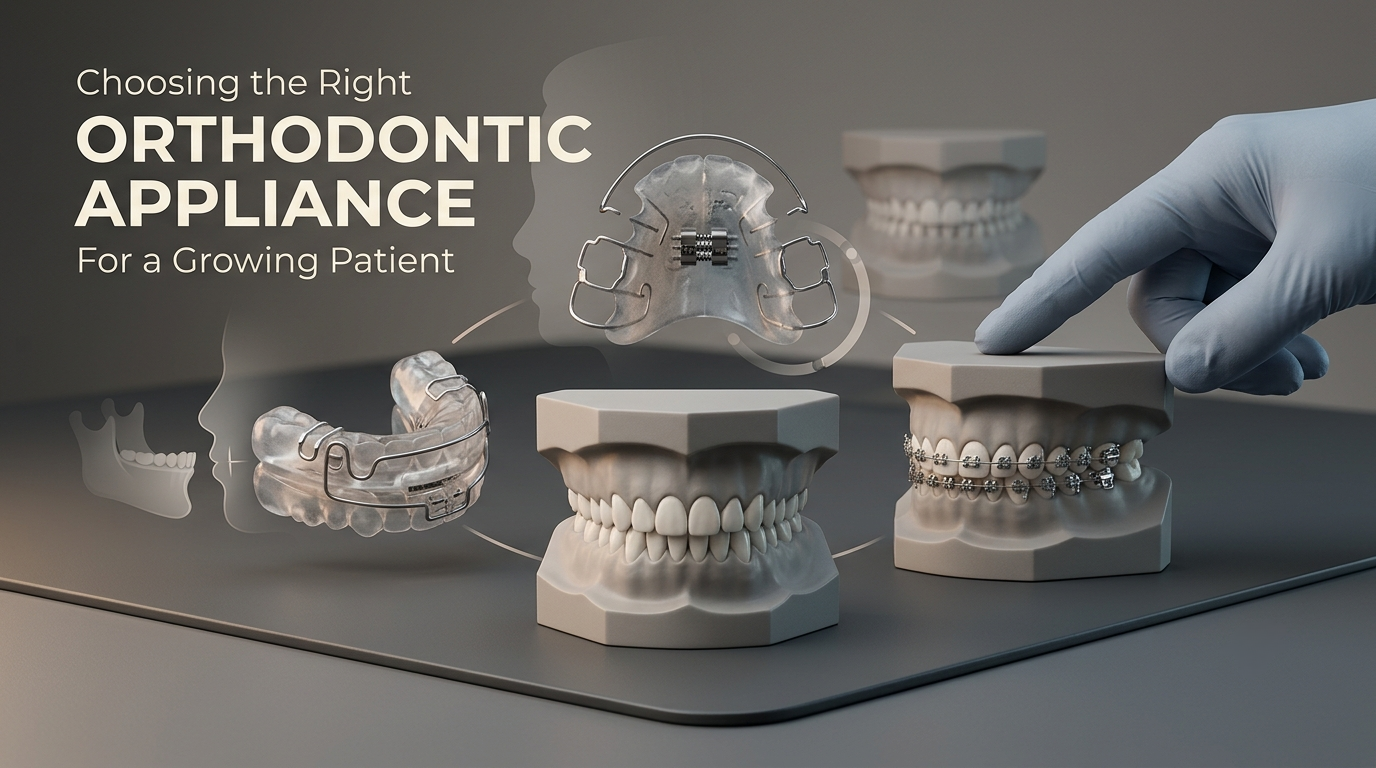
How to Choose Orthodontic Appliances for Growing Patients
How Do You Choose Between a Functional, Removable, or Fixed Orthodontic Appliance?In a growing patient, choose the appliance by the level of the problem first,
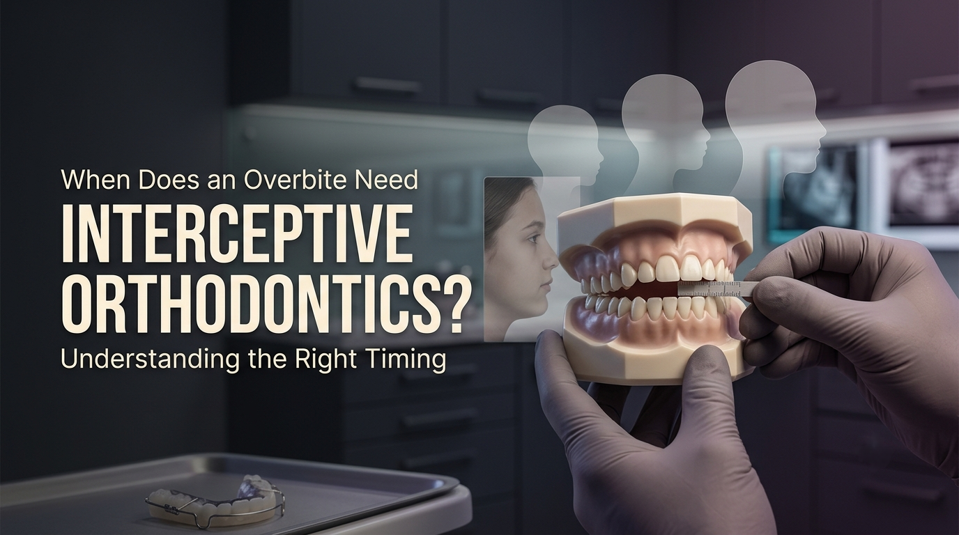
When Interceptive Orthodontics Is Needed for an Overbite
When Does an Overbite Case Call for Interceptive Orthodontics Instead of Waiting?Interceptive orthodontics is justified in an overbite case in two situations: w
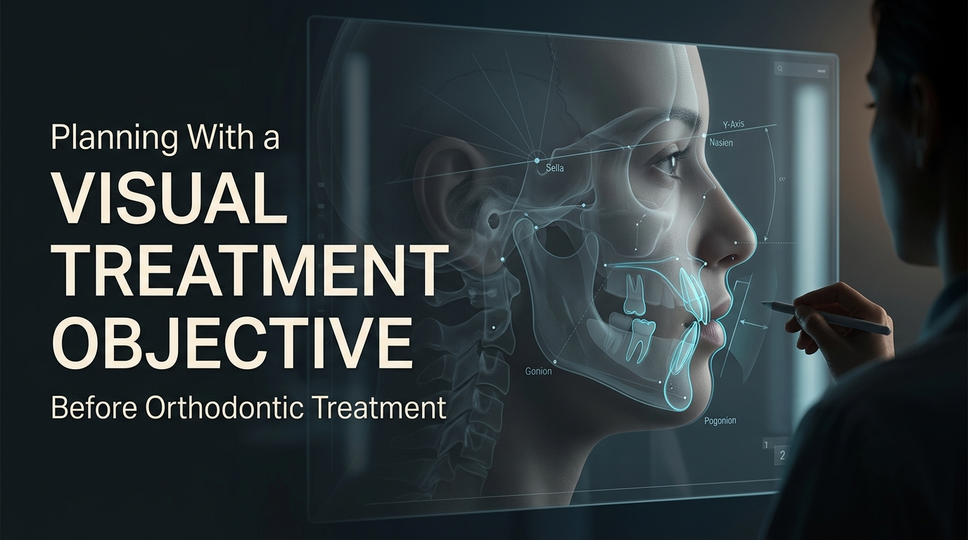
How VTO in Orthodontics Helps Plan Treatment and Outcomes
How Is a Visual Treatment Objective (VTO) Used to Plan an Orthodontic Case?A Visual Treatment Objective is used to test a plan before treatment starts. On the l