
Dr.Yukti
Dental Content Contributor
Fractured Endodontic Instrument Retrieval
A Practical Clinical Framework
A broken file in the root canal system presents a challenging clinical scenario. This guide outlines a systematic, evidence-based approach for assessing, managing, and resolving these complications effectively and predictably.
Table of Contents
Assess, Bypass, or Retrieve?
- Assess the instrument type, length, and material that has fractured.
- Evaluate the canal anatomy, including curvature and diameter at the fracture site.
- Determine the stage of cleaning and shaping when the separation occurred.
- Check the tooth's restorability and its overall role in the dental arch.
- Consider the patient's medical history and their tolerance for long procedures.
- Evaluate the operator's skill level and the availability of specialized equipment.
Clinical Decision-Making Framework
Your Instrument Retrieval Toolkit
- A dental operating microscope provides essential magnification and coaxial illumination.
- Specialized ultrasonic tips are needed to trough and vibrate the fragment.
- Staging burs, like the Gates Glidden, create a platform for access.
- Micro-forceps or loop systems can grasp the dislodged instrument.
- Small, pre-curved hand files are used to bypass the obstruction.
- A robust irrigation and suction system is needed to clear debris.
CORE COMPONENTS FOR SUCCESSFUL RETRIEVAL
A dental operating microscope is the standard of care for visualizing the fragment.
Delivers controlled micro-vibrations to loosen the instrument from canal walls.
Includes specialized forceps, loops, and extractors designed for intracanal work.
CBCT provides a 3D view to assess fragment position and surrounding anatomy.
Ultrasonic vs. Mechanical Methods
- Ultrasonics offer high precision and control in experienced hands.
- Mechanical systems often have a more defined, step-by-step protocol.
- Heat generation is a significant risk with improper ultrasonic use.
- Mechanical methods may require more coronal flaring for tube access.
- Combining both techniques can often yield the best clinical results.
- The learning curve for ultrasonic techniques is generally steeper.
Ultrasonic Technique
- Highly precise and conservative of tooth structure.
- Versatile for fragments in various canal locations.
- High risk of iatrogenic damage without proper training.
- Requires expensive equipment and specialized tips.
- Can generate excessive heat, damaging periodontal tissues.
Mechanical Systems
- More structured and predictable workflow for beginners.
- Less technique-sensitive than freehand ultrasonic use.
- Effective for fragments with an exposed coronal end.
- Can be more aggressive in removing coronal dentin.
- May not be suitable for deeply seated or curved fragments.
Post-Retrieval and Long-Term Prognosis
- Assess remaining dentin thickness to evaluate fracture risk.
- Thoroughly irrigate the canal to remove all metallic debris.
- Recapitulate the canal to ensure patency to the original working length.
- Complete chemomechanical debridement before the final obturation.
- A well-fitting coronal restoration is essential for long-term success.
- Schedule regular follow-up appointments to monitor periapical healing.
POST-RETRIEVAL PROTOCOL FOR SUCCESS
Check for any new ledges, perforations, or transportation after retrieval.
Perform a final irrigation protocol to ensure the canal is bacteria-free.
Achieve a dense, three-dimensional seal of the entire root canal system.
Place a definitive coronal restoration immediately to prevent re-contamination.
Patient Communication is Key
After a successful retrieval, clearly explain the event and positive outcome to the patient. Document the complication and its resolution in the patient's chart, including pre-operative and post-operative radiographs showing the fragment's removal and final fill.
Frequently Asked Questions
The success rate for retrieving separated instruments varies widely, generally ranging from 65% to over 90%. Success is highest for fragments located in the coronal or middle third of straight canals. The rate drops below 50% for instruments fractured in the apical third or beyond a significant canal curvature, where bypassing becomes a more viable option.
No, it is not always necessary. If an instrument fractures late in the cleaning and shaping process in a tooth with no periapical lesion, it can often be left. The prognosis is favorable if the canal system apical to the fragment was cleaned and a good seal can be achieved coronal to it. The decision requires careful case-by-case evaluation.
A broken file does not automatically condemn a tooth to failure. The primary cause of endodontic failure is the persistence of bacteria. If the canal was adequately disinfected before the fracture, or if the fragment can be bypassed and the canal sealed, the long-term prognosis can still be very good, with success rates often exceeding 80% in such cases.
Excessive dentin removal significantly weakens the root and increases the risk of vertical root fracture. While there is no absolute value, a common clinical guideline is to preserve at least 1.0 mm of circumferential dentin thickness. Using a dental operating microscope and ultrasonic tips helps minimize dentin removal to less than 0.2 mm, preserving tooth structure.
Written by
Dr.Yukti
Dental Content Contributor
Dr. Yukti Jain is a BDS-qualified dental professional and Product Specialist at Dentalkart with expertise in dental materials, equipment, and clinical innovations. Passionate about evidence-based dentistry, she creates insightful, research-driven content that simplifies complex topics and empowers dental professionals to make informed clinical and purchasing decisions.
Keep reading
Browse all →
Bioceramic Repair of Iatrogenic Root Perforations
Bioceramic Repair of Iatrogenic Root PerforationsA Modern Endodontic Solution Bioceramic materials like MTA (mineral trioxide aggregate) and Biodentine offer a

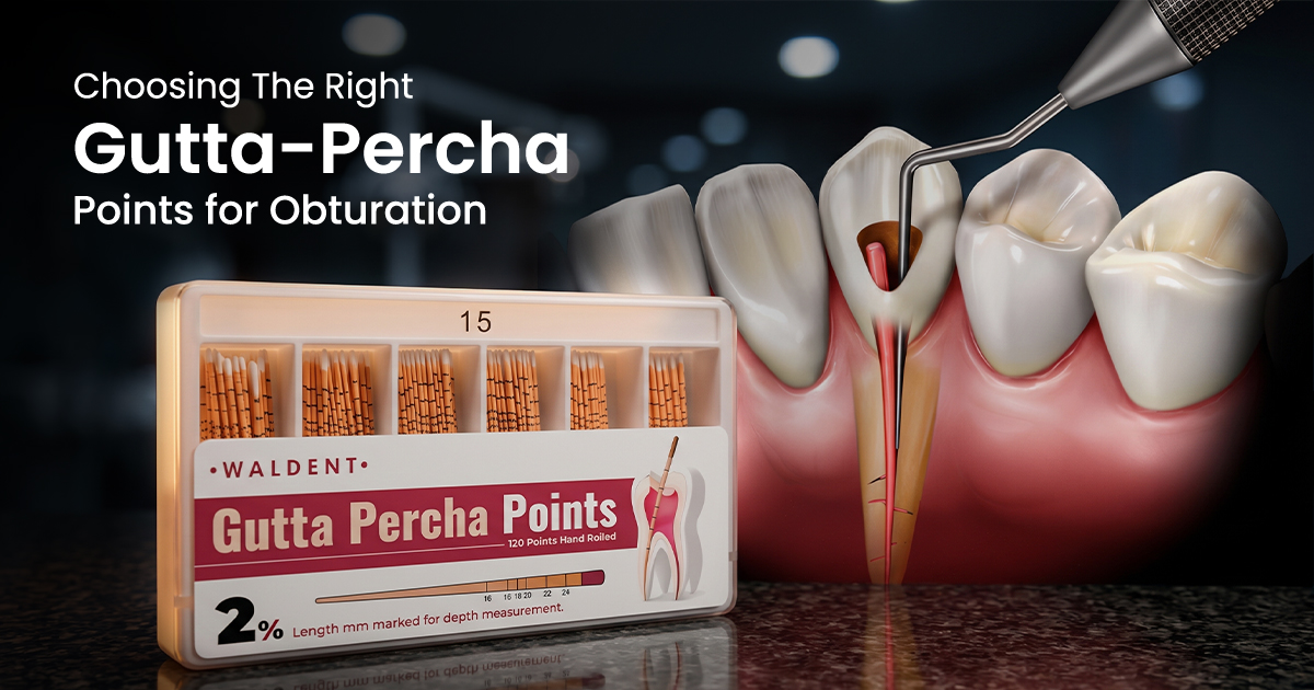
Choosing The Right Gutta-Percha Points for Obturation
Choosing The Right Gutta-Percha Points for ObturationA Clinical Selection Guide Choosing the right endodontic gutta percha point is critical for achieving a den

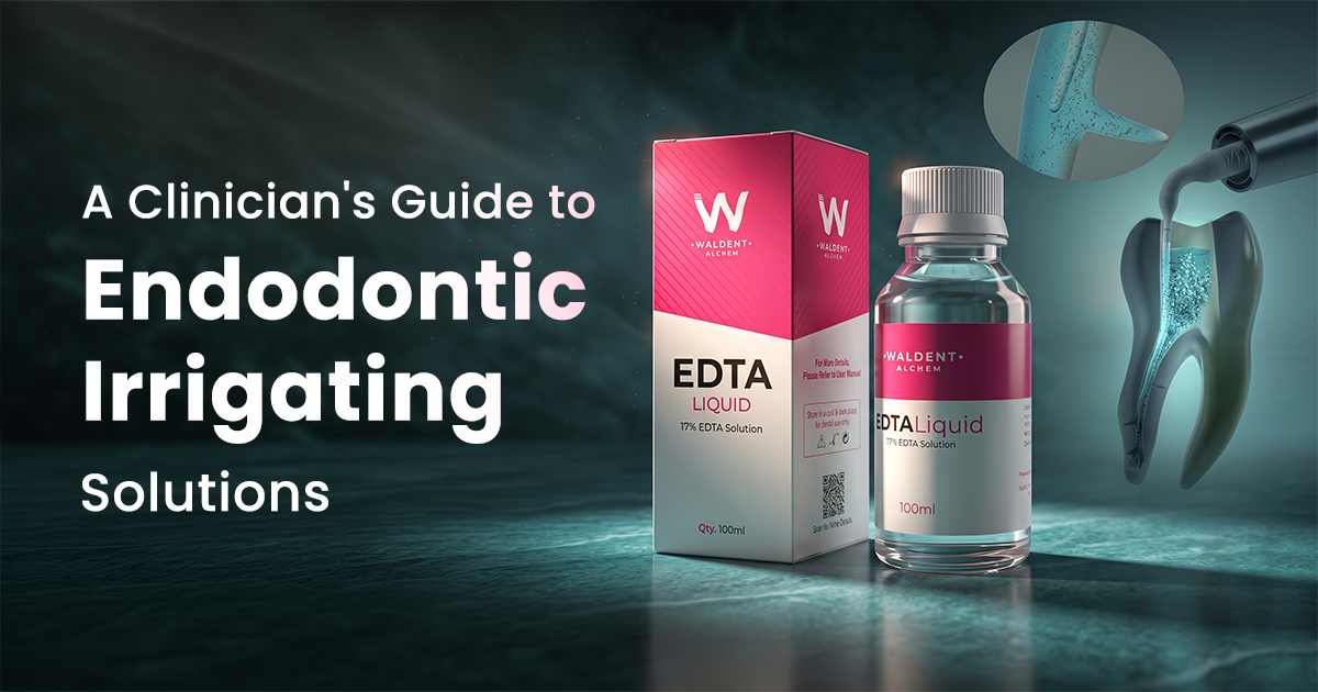
A Clinician's Guide to Endodontic Irrigating Solutions
A Clinician's Guide to Endodontic Irrigating SolutionsOptimizing Canal Disinfection Selecting the right endodontic irrigating solution involves a specific seque