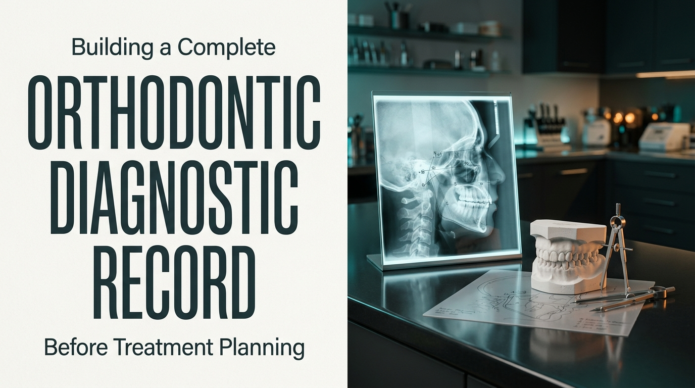
Cephalometry in Orthodontics: A Complete Diagnostic Guide

Dr. Gurjeet
Contributing Dental Clinician
How Does Cephalometry in Orthodontics Help Build a Complete Diagnostic Record?
Assemble the record from history, clinical examination, extraoral and intraoral photographs, study models or intraoral scans, space analysis and Bolton tooth-size analysis, adding radiographs such as a panoramic view and a lateral cephalogram when the clinical findings indicate them. Read each record against the others, turn the findings into one prioritised problem list, and only then set treatment objectives and choose appliances. Cephalometry in orthodontics is a powerful part of that record, but no single record, the ceph included, decides the plan on its own.
The order matters as much as the content. If you decide on aligners or extractions first and then look for supporting numbers, you will usually find them. If you build the problem list first, the plan tends to find you.
What Each Record Answers, and What It Cannot
Every component earns its place by answering a question the others cannot. That is also why none of them should dominate.
Record | Answers | Blind spot |
|---|---|---|
History and chief complaint | Patient priorities, medical status, habits, growth history | Objective severity |
Clinical examination | Functional shifts, TMJ status, periodontal support, incisor display | Skeletal source of the problem |
Extraoral (facial) photographs | Profile, symmetry, lip competence, smile arc, gingival display | Underlying jaw relationship |
Intraoral photographs | Occlusion, gingival condition, decalcification, existing restorations, baseline for consent | Root position, skeleton |
Models or intraoral scans | Arch form, crowding, spacing, curve of Spee, occlusion | Root position, skeleton |
Panoramic radiograph (when indicated) | Unerupted and missing teeth, root form, third molars, pathology | Sagittal and vertical skeletal pattern |
Lateral cephalogram (when indicated) | Jaw relationship, vertical pattern, incisor inclination, compensation | Arch length, tooth size, aesthetics |
Space analysis | Crowding in millimetres, available versus required space | Whether the skeleton tolerates expansion or proclination |
Bolton analysis | Whether the arches can fit once aligned | Which correction is clinically appropriate |
Radiographs are not taken by default. Current guidance, including the British Orthodontic Society's radiography guidelines, is based on clinical selection criteria: each film should be justified by a question it will answer for that patient. A panoramic view is usually justified at the start of treatment to check for missing, unerupted or ectopic teeth and pathology. A lateral ceph is justified when the skeletal pattern or incisor position will influence the plan, which in practice covers most cases where incisors will move anteroposteriorly.

CBCT is not a routine extra record. Reserve it for questions that 2D imaging genuinely cannot answer, such as localising an impacted canine, checking for root resorption of adjacent teeth, or assessing asymmetry in surgical cases, and justify the additional dose against the expected diagnostic gain.
Reading a Lateral Cephalogram in Layers
Jumping straight to ANB is the most common shortcut in ceph reading, and the most misleading. I work through four layers instead.
1. Check the film before the numbers
Confirm head position in the cephalostat, Frankfort horizontal orientation, teeth in occlusion, lips at rest, magnification correction and landmark clarity. Some doubling of bilateral structures such as the mandibular border is normal, because the two sides sit at different distances from the film. Marked double contours at the mandibular border or the orbits, however, signal head rotation, and every angular value built on those landmarks inherits the error. Automated and AI-assisted tracing saves time, but landmark placement still needs a human check, particularly at A point, B point and gonion, where both software and clinicians disagree most.
2. Locate the skeletal discrepancy
Steiner's mean values are SNA 82°, SNB 80° and ANB 2°. The "±2°" often quoted alongside them is narrower than the spread most population studies report: standard deviations of roughly 3° for SNA and SNB are more typical. Treat these as population references, not treatment targets. They also shift with ethnicity, age and sex, and Indian population norms differ from the original Caucasian sample.
ANB alone cannot tell you where the problem sits. Consider two hypothetical patients, both with an ANB of 5°:
Measure | Patient A | Patient B |
|---|---|---|
SNA | 87° | 82° |
SNB | 82° | 77° |
ANB | 5° | 5° |
Interpretation | Prominent maxilla, mandible within the normal range | Maxilla within the normal range, retrusive mandible |
Direction of treatment | Maxillary restraint may be considered | Candidate for mandibular advancement during growth |

The same ANB, two different problems. ANB is also distorted by the position of nasion and by jaw rotation, so confirm it with a second sagittal indicator. The Wits appraisal (Jacobson) measures the distance between the projections of A and B points on the functional occlusal plane, with mean values of about 0 mm in females and −1 mm in males.
3. Establish the vertical pattern
Mandibular plane angle, palatal plane inclination and the ratio of posterior to anterior face height shape anchorage planning, tolerance for molar extrusion, bite-opening mechanics and long-term stability. A high-angle and a low-angle Class II with identical sagittal values need different mechanics. Class II elastics and cervical headgear, both of which tend to extrude molars, suit the high-angle patient poorly.
4. Read incisor inclination as compensation
Incisors frequently tip to disguise the skeletal pattern. Proclined lower incisors in a Class II case and retroclined lower incisors in a Class III case are classic dentoalveolar compensations. The ceph tells you where the tooth sits. Whether it can safely move further depends on symphysis width, labial gingival thickness, lip support and the planned overjet, which is why the ceph must be read alongside the clinical examination. If surgery is planned, these compensations are usually removed before the operation, deliberately worsening the occlusion so the jaws can be repositioned fully.
Bolton Analysis: Check Arch Fit Before Space Closure
Bolton analysis compares the mesiodistal widths of the mandibular and maxillary teeth:
Anterior ratio: the combined width of the lower six anterior teeth as a percentage of the upper six
Overall ratio: the same comparison across all twelve teeth in each arch, first molar to first molar
Bolton's original means are 77.2% (anterior) and 91.3% (overall). Population studies report slightly different means, so use these as references rather than fixed ideals.
Worked example (hypothetical measurements)
Measurement | Value |
|---|---|
Sum of upper 6 anteriors | 47.0 mm |
Sum of lower 6 anteriors | 38.0 mm |
Anterior ratio | 80.9% |
Ideal lower 6 for this upper arch | 36.3 mm |
Mandibular anterior excess | 1.7 mm |

A 1.7 mm mandibular excess means that even with perfectly aligned arches, achieving ideal overjet, overbite and Class I canines will be difficult. Discrepancies above about 1.5 mm are commonly cited as clinically significant (Proffit and colleagues). How you respond depends on which teeth carry the excess and the planned finish: interproximal enamel reduction in the lower labial segment, accepting slightly reduced overbite or increased overjet, or adding width in the upper arch with composite build-ups where small or peg-shaped laterals are the cause.
The real lesson is timing. Found at the records stage, a Bolton discrepancy shapes the plan. Found after space closure, it means reopening spaces or compromising the finish.
Converting Findings Into a Problem List
Translate every finding into plain clinical language, group it by domain and prioritise. A finding only earns its place if it changes diagnosis, prognosis, mechanics, timing or consent.
Domain | Example finding | Planning consequence |
|---|---|---|
Facial | Convex profile, lip strain at rest | Limits incisor proclination; extraction may improve lip competence |
Skeletal (sagittal) | Class II, mandibular source | Growth modification if growth remains |
Vertical | Increased mandibular plane angle | Avoid molar extrusion; consider vertical anchorage control |
Transverse | Unilateral posterior crossbite with a narrow maxilla and a functional shift | Maxillary expansion, timed while the midpalatal suture still responds |
Space and incisors | 6 mm lower crowding, proclined lower incisors | Further proclination unlikely to be stable |
Tooth size | 1.7 mm mandibular anterior excess | Plan IPR or adjust finishing targets |
Growth | Pre-pubertal stage | Functional appliance timing is favourable |
With the list in place, objectives follow, and appliances follow the objectives. For the materials used at this stage, Dentalkart's Diagnosis and Treatment Planning category stocks orthodontic diagnostic materials and accessories.
When the Records Disagree
Inconsistency between records is itself a finding. A profile that looks Class III with a Class I ceph, landmarks that cannot be located reliably, a severe skeletal discrepancy, or a plan that depends on heavy dentoalveolar compensation all justify a second opinion, a repeat radiograph or a surgical consultation. Two-dimensional cephalometry has known limits with asymmetry and superimposed bilateral structures, and that is exactly where it should be supplemented rather than trusted.
Frequently Asked Questions
Chronological age is a weak guide. The cervical vertebral maturation (CVM) method uses the lateral cephalogram you already have, classifying C2 to C4 morphology into six stages, with the pubertal growth peak expected between CS3 and CS4 (Baccetti and colleagues, 2005). Functional appliance treatment timed around this peak tends to produce more skeletal response. Be aware that CVM staging varies between observers, particularly in the CS2 to CS4 range; if the stage is borderline and the decision depends on it, have a second clinician stage the film or re-stage at the next indicated radiograph. A hand-wrist radiograph is now reserved for the occasional case where the CVM stage remains genuinely unclear and the treatment decision depends on it.
For full-arch measurement of tooth widths and arch length, studies generally report digital models within clinically acceptable agreement of plaster casts. The practical gains are storage, instant measurement tools and direct use in aligner and indirect bonding workflows. Plaster remains useful where scanning access is limited or a physical articulator mounting is planned.
A progress cephalogram is justified when it will change the next decision: confirming incisor position before finishing a camouflage case, assessing response to growth modification, or re-planning before surgery. Routine repeat imaging without a specific question adds exposure without adding information.
Written by
Dr. Gurjeet
Contributing Dental Clinician
Dr. Gurjeet is a practising dental clinician contributing to Dentalkart Blogs, sharing chair-side experience, treatment protocols, and equipment insights for the wider dental community.
Keep reading
Browse all →
How to Choose Orthodontic Appliances for Growing Patients
How Do You Choose Between a Functional, Removable, or Fixed Orthodontic Appliance?In a growing patient, choose the appliance by the level of the problem first,
When Interceptive Orthodontics Is Needed for an Overbite
When Does an Overbite Case Call for Interceptive Orthodontics Instead of Waiting?Interceptive orthodontics is justified in an overbite case in two situations: w
How VTO in Orthodontics Helps Plan Treatment and Outcomes
How Is a Visual Treatment Objective (VTO) Used to Plan an Orthodontic Case?A Visual Treatment Objective is used to test a plan before treatment starts. On the l