
Dr. Bhavishya Arora
Chief Dental Editor
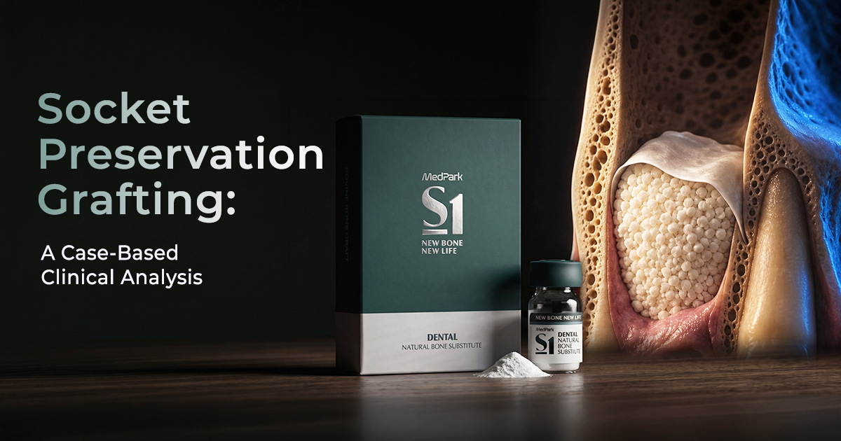
Socket Preservation Grafting: A Case-Based Clinical Analysis
Selecting Materials for Ridge Integrity
Socket preservation — also called alveolar ridge preservation — involves placing a dental bone graft material into an extraction socket to minimize alveolar ridge resorption. This is clinically critical for preserving bone volume and architecture, ensuring predictable future implant placement and superior aesthetic outcomes in implant dentistry.
Table of Contents
Grafting an Anterior Maxillary Socket
In the anterior maxilla, socket preservation prioritizes maintaining the buccal bone plate to support soft tissue aesthetics for a future implant crown. The primary goal is volumetric stability. An atraumatic extraction is mandatory to preserve the delicate socket walls, especially the buccal plate, which is often less than 1 mm thick.
A slow-resorbing graft material is typically preferred to act as a long-term scaffold, preventing ridge collapse as native bone remodels. This is often a cortico-cancellous xenograft particulate. The graft must be packed gently, avoiding excessive pressure that could compromise the thin bony walls or local blood supply, which is critical for successful angiogenesis and new bone formation. Proper material selection from a range of periodontic products is the first step toward a successful outcome.
- Material Choice: A composite graft (e.g., 70% xenograft, 30% alloplast) can provide both long-term scaffolding and predictable handling.
- Buccal Plate Integrity: If the buccal plate is compromised or thinner than 1 mm post-extraction, a barrier membrane is essential.
- Graft Placement: Condense the graft material incrementally, ensuring it fills the socket up to the crestal bone level but not beyond.
- Suturing Technique: Achieve tension-free primary closure using criss-cross or horizontal mattress sutures to contain the graft and protect it from the oral environment.
Material Selection in the Aesthetic Zone
Xenograft vs Alloplast for Molar Sockets
For posterior molar sockets, the choice between a xenograft and an alloplast depends on the clinician's philosophy regarding resorption rates and the desired healing timeline. Xenografts, typically bovine-derived, are osteoconductive and resorb very slowly, providing excellent long-term volume maintenance which is advantageous in large defects.
Alloplasts, which are synthetic materials like hydroxyapatite (HA) or beta-tricalcium phosphate (β-TCP), offer a completely sterile, non-immunogenic alternative with variable resorption profiles. A pure β-TCP alloplast will resorb relatively quickly (6-12 months) as it is replaced by host bone, whereas a biphasic calcium phosphate (HA/β-TCP) provides both a stable scaffold and a resorbable component. The choice impacts the quality of bone available for primary implant stability later.
- Xenografts: Excellent for maintaining ridge width and height over time, making them ideal for delayed implant placement.
- Alloplasts (β-TCP): Suited for cases where faster replacement by vital bone is prioritized, potentially allowing for slightly earlier implant placement.
- Alloplasts (Biphasic): Offer a balanced approach, with the HA component providing structural integrity while the β-TCP portion remodels.
- Particle Size: For large molar sockets, a larger particle size (1.0-2.0 mm) helps maintain space and prevent rapid resorption.
Xenograft (Bovine)
- Excellent volume stability due to very slow resorption.
- Extensive clinical documentation and predictable outcomes.
- Osteoconductive scaffold structure mimics human bone.
- Theoretical risk of disease transmission (extremely low).
- Slower replacement with host vital bone.
Alloplast (Synthetic)
- Zero risk of biological contamination or immune response.
- Controlled resorption rates (fast with β-TCP, slow with HA).
- Consistent product quality and availability.
- Lacks biologic induction factors; purely osteoconductive.
- Some formulations may show incomplete resorption.
Choosing a Barrier Membrane for Grafts
The primary function of a barrier membrane in socket grafting is to prevent the apical migration of epithelial cells and the infiltration of gingival connective tissue into the graft, allowing slower-growing osteogenic cells to populate the scaffold. The choice between resorbable and non-resorbable membranes is dictated by the defect size and need for primary closure.
Resorbable collagen membranes are the most common choice for routine socket preservation due to their biocompatibility and ease of use, as they do not require a second surgical procedure for removal. Their resorption time varies from 4 to 24 weeks depending on the degree of cross-linking. Non-resorbable membranes, like dense-PTFE (d-PTFE), provide extended barrier function and are indicated for larger defects or when primary closure is not achievable, as they can be left intentionally exposed.
- Collagen Membranes: Ideal for contained defects where primary closure is easily achieved. Select a longer-lasting cross-linked membrane for larger grafts.
- d-PTFE Membranes: Use for large buccal defects or when flap advancement is limited. Requires removal after 4-6 weeks.
- Membrane Fixation: For most socket grafts, the membrane can be tucked under the buccal and lingual flaps for stability. Tacks or sutures are only needed for large GBR procedures.
- Hydration: Always hydrate collagen membranes in sterile saline before placement to make them pliable and prevent tearing.
| Feature | Resorbable (Collagen) | Non-Resorbable (d-PTFE) |
|---|---|---|
| Barrier Function Time | 4-24 weeks | Until removed (stable) best |
| Second Surgery | No | Yes (for removal) |
| Handling | Pliable when hydrated, can tear | Stiffer, excellent space-making |
| Exposure Management | Risk of rapid degradation | Can be left exposed value |
| Indication | Contained sockets, GBR with closure | Non-contained defects, compromised closure |
Assessing Graft Healing for Implant Placement
Graft healing and readiness for implant placement are assessed after a healing period of 4 to 6 months, depending on the material used and the size of the original socket. The evaluation is both clinical and radiographic. Clinically, the healed ridge should feel firm to palpation, indicating good bone fill, and display a healthy band of keratinized gingiva.
Radiographically, a Cone Beam CT (CBCT) is the gold standard for assessment. It allows for precise measurement of the final ridge dimensions (bucco-lingual width and height) and evaluation of bone density. The grafted bone should appear well-integrated with the native bone, without clear lines of demarcation or radiolucent areas that would suggest fibrous encapsulation or incomplete healing. A minimum of 5 mm of bone width is generally required for standard implant placement.
- Healing Period: 4 months for alloplasts/allografts; 5-6 months for xenografts.
- Clinical Check: Palpate the ridge to ensure it is hard and non-compressible. Check soft tissue quality and volume.
- Radiographic Check (CBCT): Verify ridge width >5-6 mm and height. Assess bone quality and ensure uniform radiopacity.
- Implant Timing: Proceed with implant placement only when both clinical and radiographic signs confirm successful, mature bone formation capable of providing primary stability.
Day 0: Extraction & Grafting
Atraumatic extraction, socket degranulation, placement of dental bone graft material and membrane, and tension-free primary closure.
Week 1-2: Soft Tissue Healing
Initial wound closure and epithelialization. Suture removal typically at 10-14 days. Patient maintains soft diet and meticulous hygiene.
Month 1-3: Initial Bone Formation
Angiogenesis and osteoid deposition begin within the graft scaffold. The graft is gradually resorbed and replaced by woven bone.
Month 4-6: Bone Maturation & Assessment
Woven bone remodels into mature lamellar bone. Perform clinical and CBCT evaluation to confirm ridge dimensions and density for implant placement.
Frequently Asked Questions
The primary role of a dental bone graft material in socket preservation is to act as a scaffold, or framework, that prevents soft tissue collapse and supports the formation of new bone. It maintains the three-dimensional volume of the alveolar ridge after tooth extraction, which is essential for ensuring sufficient bone is available for the future placement of a dental implant.
The main difference lies in their mechanism of action. Osteoconductive materials, like most xenografts and alloplasts, provide a passive scaffold that allows new bone from the patient to grow into it. Osteoinductive materials, such as autografts or allografts containing bone morphogenetic proteins (BMPs), actively stimulate undifferentiated cells to become bone-forming osteoblasts, thereby inducing new bone formation.
For a socket with a buccal wall dehiscence, a slow-resorbing, volume-stable graft like a xenograft is generally preferred. This must be combined with a durable, space-making barrier membrane (Guided Bone Regeneration) to contain the graft and prevent soft tissue invasion. The membrane ensures the space is maintained long enough for bone cells to regenerate the missing wall.
Premature membrane exposure is most commonly caused by a lack of tension-free primary closure of the soft tissue flap over the graft site. Other causes include excessive graft volume creating pressure on the flap, poor suturing technique, or patient-related factors like smoking or poor oral hygiene, all of which can compromise blood supply and lead to tissue necrosis and recession.
The standard waiting period for implant placement after socket preservation is 4 to 6 months. This allows sufficient time for the graft material to be resorbed and replaced by mature, vital host bone capable of providing adequate primary stability for the implant. The exact timing depends on the specific dental bone graft material used, the size of the socket, and the patient's healing capacity.
Written by
Dr. Bhavishya Arora
BDS, MDSChief Dental Editor
A contributing clinician at Dentalkart Blogs, Dr. Bhavishya is a compassionate, detail-driven dentist known for thoughtful patient care, clear communication, and a calm chair-side manner that puts patients at ease. Beyond the clinic, she leads product content and category initiatives at Dentalkart India's largest dental marketplace where she bridges clinical understanding with commercial insight to help dentists make better-informed product choices.
Keep reading
Browse all →
Indications for Periodontal Dressing Placement
Indications for Periodontal Dressing PlacementA Clinical Case Selection Guide A periodontal dressing material is a protective pack placed over a surgical site t

Indications for Periodontal Fiber Splinting
Indications for Periodontal Fiber SplintingA Clinical Case Selection Guide Periodontal splinting is indicated for stabilizing teeth with increasing mobility (Mi
Preventing Burnished Calculus with Gracey Curettes
Preventing Burnished Calculus with Gracey CurettesA Subgingival Scaling Protocol Burnished calculus is a residual deposit smoothed by an incorrectly used curett