
Dr. Prerna Sahrawat
Contributing Dental Clinician
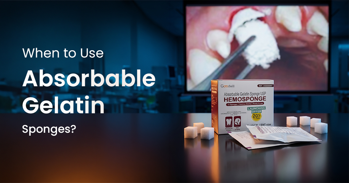
When to Use Absorbable Gelatin Sponges?
Clinical Hemostasis Case Selection
An absorbable dental hemostatic sponge is a sterile, porcine-derived gelatin foam used to control post-operative bleeding by providing a physical matrix for clot formation. Its primary clinical value is in managing persistent oozing from extraction sockets where simple pressure is insufficient, especially in anticoagulated patients.
Table of Contents
Clinical Indications for Gelatin Sponges
A gelatin sponge is indicated for capillary or venous bleeding in extraction sockets and surgical sites where primary closure is not feasible or where underlying patient factors complicate hemostasis. Its porous structure absorbs approximately 40-50 times its weight in blood, creating a large surface area that accelerates the clotting cascade through platelet aggregation and activation. This makes it particularly effective for achieving a stable clot that conforms to the socket's architecture.
Use is warranted in cases beyond routine extractions, providing a scaffold for healing and preventing hematoma formation in deeper bony defects. For patients on anticoagulants like warfarin or newer direct oral anticoagulants (DOACs), it provides a crucial local measure to achieve hemostasis without altering their systemic medication regimen, provided their INR is within the therapeutic range (typically 2.0-3.5).
- Anticoagulated Patients: Local hemostasis for patients on warfarin, clopidogrel, or DOACs with therapeutic INR levels.
- Deep Bony Defects: To manage bleeding and prevent hematoma after cyst enucleation or apicoectomy.
- Multiple Extractions: When widespread socket oozing is anticipated and difficult to control with pressure alone.
- Friable Tissue: In cases where suturing is difficult due to poor tissue quality, such as in irradiated patients or those with certain systemic diseases.
Case Selection Guide for Gelatin Sponge Use
Gelatin Sponge Contraindications and Cautions
Gelatin sponges should not be used in the presence of active infection or in grossly contaminated wounds. The sponge can act as a nidus for bacterial growth, potentially leading to a post-operative abscess and delaying healing. They are also ineffective for controlling pulsatile, arterial hemorrhage; such bleeding requires direct ligation or cauterization of the vessel.
Avoid leaving the sponge to protrude from the socket, as this can cause patient discomfort and promote dislodgement. Furthermore, do not use it to close skin incisions, as its presence can interfere with the epithelialization process and normal wound edge healing. It is a hemostatic agent for cavities and spaces, not a primary closure material.
- Known Allergy: Contraindicated in patients with a known allergy to porcine collagen or gelatin.
- Frank Pus: Do not place in sockets with evidence of purulent exudate or an established abscess.
- Arterial Bleeding: Ineffective for high-pressure arterial bleeds which require definitive vascular control.
- Intravascular Compartments: Never leave within blood vessels, as it can cause embolization.
Avoid Over-Packing the Socket
Do not compress the gelatin sponge into a hard, dense plug. Over-packing can exert excessive pressure on the underlying bone and neurovascular bundles, leading to ischemia, post-operative pain, and delayed healing. The sponge should fit passively within the socket.
Properly Placing a Gelatin Sponge
Correct placement technique is critical for the efficacy of a gelatin sponge. The sponge should be trimmed to fit the dimensions of the socket; it should not be forced or packed tightly. If desired, it can be pre-moistened with sterile saline or thrombin solution, though it is often placed dry to maximize its absorptive capacity for blood within the socket.
Once inserted, it should fill the socket space without extending significantly above the alveolar crest. Gentle pressure is then applied with a sterile gauze pack for 2-5 minutes. This ensures intimate contact between the sponge, blood, and socket walls, which is necessary for initiating the clotting process. In many cases, especially with anticoagulated patients, placing a suture over the socket helps retain the sponge and stabilize the clot.
- Socket Debridement: Ensure the socket is free of bone fragments, granulation tissue, and debris.
- Passive Fit: Cut the sponge to size. It should be slightly smaller than the socket to allow for expansion as it absorbs blood.
- Suture Retention: A figure-of-eight or horizontal mattress suture is recommended to secure the sponge.
- Patient Instructions: Advise patients to avoid vigorous rinsing or spitting for 24 hours to prevent dislodging the clot.
Step 1: Isolate and Prepare
After extraction, irrigate the socket with sterile saline to remove debris. Achieve clear visibility and gently suction excess fluid, ensuring the socket walls are clean.
Step 2: Trim and Adapt
Using sterile scissors, trim the gelatin sponge to the approximate size and depth of the socket. For multi-rooted teeth, consider using smaller pieces for each root socket.
Step 3: Insert and Secure
Gently insert the trimmed sponge into the socket until it reaches the apex. Place a gauze pack over the site and have the patient bite with firm, steady pressure for 2-5 minutes. Secure with a suture if needed.
Gelatin Sponge vs. Other Hemostatic Agents
The choice between a gelatin sponge and other local hemostatic agents depends on the clinical situation, handling characteristics, and cost. While gelatin provides an excellent physical matrix for clot formation, other materials offer different properties. Oxidized regenerated cellulose (ORC), for example, provides hemostasis and has a low pH, which imparts a bactericidal effect but can slightly delay epithelial healing.
Collagen plugs and sponges are derived from bovine collagen and provide a more robust structural scaffold, making them ideal for socket preservation procedures ahead of implant placement. They promote tissue ingrowth more actively than gelatin. However, for pure hemostasis in a standard extraction, the rapid absorption and cost-effectiveness of a dental hemostatic sponge make it a frequent first-line choice in high-volume Indian dental practices.
- Handling: Gelatin sponges are pliable and can be cut easily, but may become flimsy when saturated. Collagen plugs are firmer and hold their shape better.
- Mechanism: All act as scaffolds, but ORC also has a chemical hemostatic effect and is bactericidal.
- Resorption Time: Gelatin typically resorbs in 4-6 weeks. Collagen resorption can take longer, which is advantageous for grafting.
| Feature | Gelatin Sponge | Collagen Plug | Oxidized Cellulose (ORC) |
|---|---|---|---|
| Primary Material | Porcine Gelatin | Bovine Collagen (Type I) | Regenerated Cellulose |
| Mechanism | Physical matrix, platelet aggregation | Platelet aggregation, chemotactic premium | Physical matrix, low pH |
| Resorption Time | 4–6 weeks | 4–8 weeks (variable) | 1–2 weeks |
| Handling | Soft, pliable, fragments easily | Firm, holds shape | Fabric-like, can be layered value |
| Primary Use Case | General hemostasis | Socket preservation, grafting | Hemostasis with bactericidal need |
Frequently Asked Questions
A dental hemostatic sponge is fabricated from purified porcine skin gelatin, which is whipped into a porous foam, baked, and sterilized. This creates a biocompatible, absorbable material that provides a physical scaffold to promote platelet aggregation and accelerate the formation of a stable blood clot within a surgical site or extraction socket.
The primary difference lies in their material and structure. A gelatin sponge is made of denatured collagen (gelatin) and has a random, porous foam structure ideal for absorbing blood. A collagen plug is made of intact, non-denatured bovine collagen, providing a firmer, more organized scaffold that is more conducive to tissue ingrowth, making it preferable for socket preservation procedures.
If a gelatin sponge dislodges within the first 24-48 hours, it can lead to secondary bleeding. The patient should be instructed to apply firm pressure with a clean gauze pack for 20-30 minutes. If bleeding persists, they must return to the clinic. The socket may need to be irrigated and a new hemostatic agent placed.
No, a gelatin sponge is not a treatment for alveolar osteitis (dry socket). A dry socket is a condition of delayed healing, often with a lost blood clot, not active bleeding. Treatment involves irrigating the socket to remove debris and placing a medicated dressing, like zinc oxide eugenol paste, to obtund pain and promote healing.
An absorbable gelatin sponge is typically resorbed by the body through enzymatic action and phagocytosis within 4 to 6 weeks. It does not need to be removed. During this time, it provides a stable scaffold which is gradually replaced by granulation tissue and, eventually, new bone as the extraction socket heals.
Written by
Dr. Prerna Sahrawat
Contributing Dental Clinician
Dr. Prerna Sahrawat is a dentist with over 4 years of experience in clinical dentistry and dental content writing. She is passionate about simplifying complex dental concepts into practical, evidence-based content that helps dentists make informed clinical and purchasing decisions while staying updated with the latest advancements in dentistry.
Keep reading
Browse all →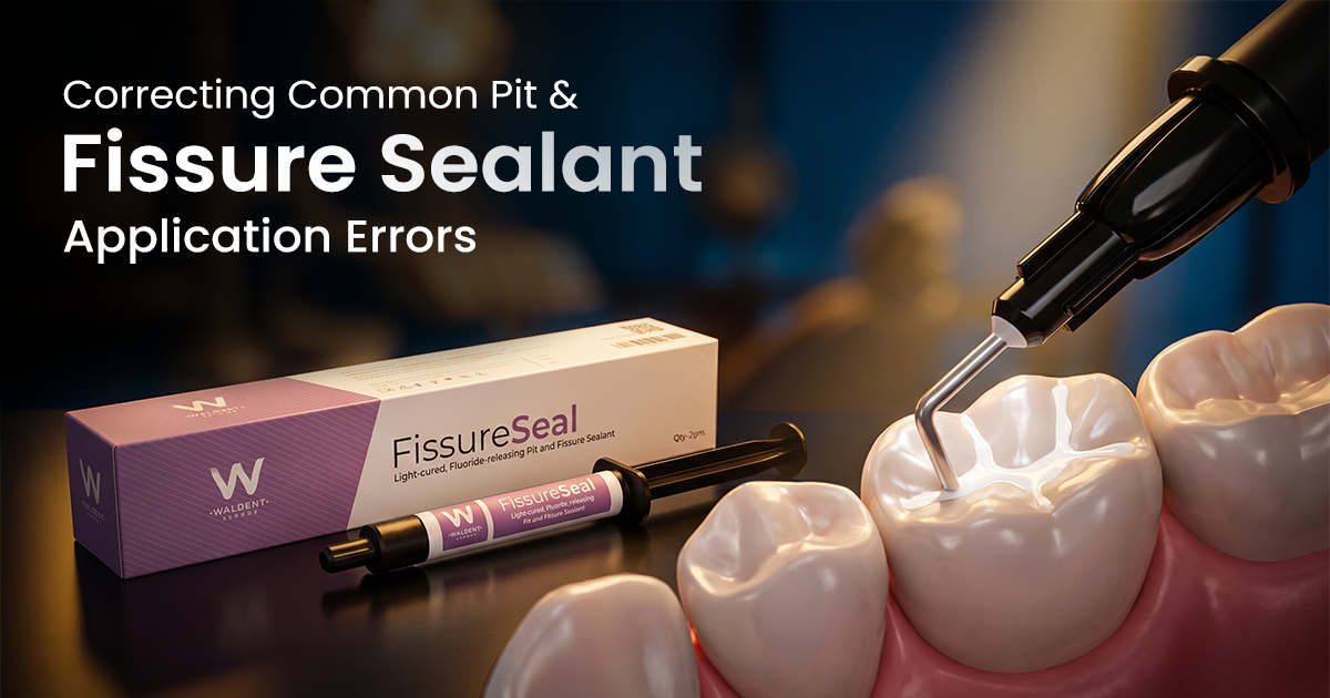
Correcting Common Pit & Fissure Sealant Application Errors
Correcting Common Pit & Fissure Sealant Application ErrorsAn Evidence-Based Guide Successful pit and fissure sealant application is technique-sensitive, and


Dental RVG Sensor Quality Assurance Protocol
Dental RVG Sensor Quality Assurance ProtocolA Clinical Calibration Guide A dental RVG (radiovisiography) sensor quality assurance protocol is a set of routine t
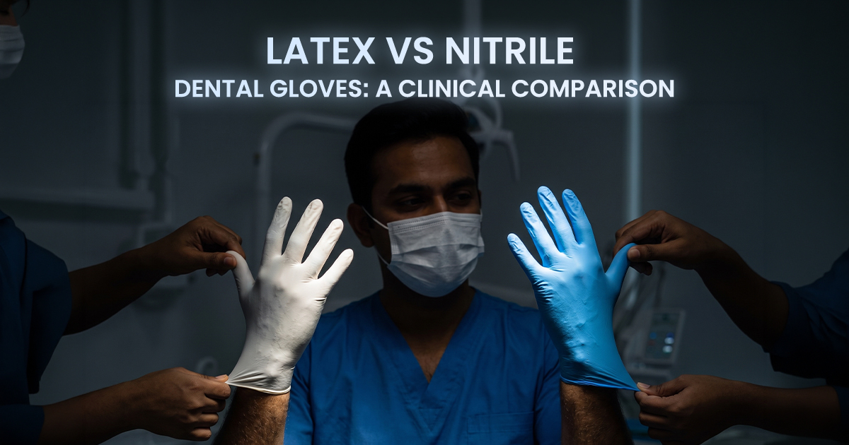
Latex vs Nitrile Dental Gloves: A Clinical Comparison
Latex vs Nitrile Dental Gloves: A Clinical ComparisonYour Clinical Selection Guide The choice between latex and nitrile dental examination gloves balances latex